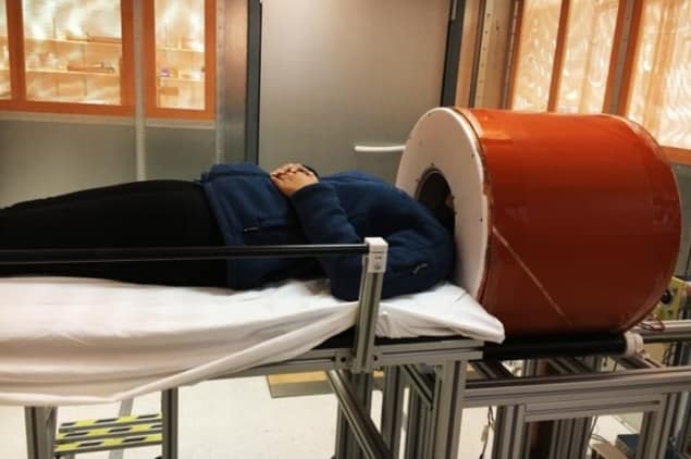
MRI is the standard modality for assessing neurological disorders, due to its ability to image intracranial anatomy with unparalleled soft-tissue contrast. Conventional high-field MRI scanners, however, are costly, immobile and require dedicated power and cooling infrastructure. As such, MRI is unavailable to critically ill patients who cannot be safely transported to the scanner or patients in low-resource settings.
A low-cost, portable brain MRI scanner could expand access to MR neuroimaging, as well as enabling point-of-care diagnostics for neurological emergencies. With this aim, researchers at Massachusetts General Hospital/Harvard Medical School are developing a portable scanner based on a compact, lightweight permanent magnet. Writing in Nature Biomedical Engineering, the researchers describe the design and testing of their prototype system.
“There are cases where MR brain imaging would be diagnostically useful, but it is not feasible because of the logistical burden and cost,” says first author Clarissa Cooley. “To address this, we wanted to develop a truly portable MRI brain scanner that could be used in new locations, like a patient’s bedside or rural clinic. Our design is meant to be a very accessible MRI option for detecting brain abnormalities that are visible at a lower field and lower resolution.”
Optimized design
The team’s portable MRI scanner is based around four key design points. First, by creating a dedicated brain scanner with a small-diameter bore that fits around the head, rather than a full-body system, the scanner size and cost can be reduced.
At the heart of the scanner is a permanent magnet made from an array of neodymium (NdFeB) rare-earth magnets that generate an 80 mT static field. Unlike the bulky superconducting magnets used in conventional MRI systems, or previously used electromagnets, the permanent magnet does not require external power or cryogenic cooling.
Arranging the magnet segments in an optimized Halbach cylinder configuration creates a transverse field inside the magnet and zero field outside the magnet. This intrinsic self-shielding is ideal for portable applications where stray fields could pose safety hazards. The constructed magnet assembly is 49 cm long, with an outer diameter of 57 cm and 27 cm bore opening.
The third design factor is that, rather than designing a homogeneous magnet, the team shaped its magnetic field variation into a built-in field gradient (of 7.6 mT/m) for readout encoding. This reduces magnet size and cost, and eliminates the need for a traditional readout gradient coil, lowering the acoustic noise, power and cooling requirements. With the built-in field variation used for image encoding in the x dimension, switchable gradient coils provide phase encoding in the y and z directions. The RF transmit/receive coil is incorporated into a compact helmet.
Finally, the researchers used advanced reconstruction techniques to correct for image distortions that arise from the non-linear field gradients used to encode the image. “Our image reconstruction method utilizes measured magnetic field maps to correct for these distortions,” Cooley explains.
Proof-of-principle
The prototype scanner – including the 122-kg magnet, coils, amplifiers, console and cart – weighs approximately 230 kg. Replacing the general-purpose console, amplifiers and cart with custom lightweight designs could reduce this to roughly 160 kg, the team notes. With no refrigeration systems for a superconducting magnet, power requirements are low, enabling the scanner to be operated from a standard power outlet.
Cooley and colleagues used their prototype scanner to record MR images from three healthy volunteers. The scanner successfully generated T1-weighted, T2-weighted and proton density-weighted brain images – standard brain scans routinely used for detection, diagnosis and monitoring of clinically important brain pathology. Each image was acquired in roughly 10 min and had a spatial resolution of 2.2 × 1.3 × 6.8 mm.
Although the scanner’s spatial resolution and sensitivity are both lower than that of a high-field MRI, the researchers emphasize that its performance is sufficient to detect and characterize serious intracranial processes, such as haemorrhage, hydrocephalus, infarction and mass lesions. Preliminary work also suggests that diffusion-weighted imaging, which is critical to applications such as acute stroke detection, should also be possible.

Portable scanner could boost point-of-care brain MRI
These initial images were acquired in an RF shielded room to eliminate external electromagnetic interference (EMI). “For true portable imaging, we are integrating EMI detectors into our scanner for EMI mitigation,” says Cooley. “This will greatly increase the image quality when our scanner is operated at the point-of-care.”
“We are also excited to begin work on a point-of-care MRI scanner specially designed for neonatal patients in the [neonatal intensive care unit] NICU,” she tells Physics World. “The transport and scanning of sick neonates is logistically very difficult and can be dangerous. The availability of a bedside MRI scanner in the NICU could have tremendous benefits for diagnostics and monitoring of neonatal brain injury.”


