
Modern imaging techniques have yielded fresh insights into how insect larvae use powerful suction organs to move around fast-moving alpine waterways. The work, by researchers in the UK and Germany, reveals the internal structure of the organs in three dimensions and highlights features that could aid the design of bio-inspired tools for attaching to smooth and rough surfaces in wet and dry conditions.
The aquatic larvae of net-winged midges beat all records of insect attachment strength. The six suction cups on the bottom of their streamlined bodies attach so tightly that forces greater than 600 times their body weight are needed to dislodge them. This allows the larvae to graze on algae in alpine streams and rivers that can flow as fast as three metres per second.
“The force of the river water where the larvae live is absolutely enormous, and they use their suction organs to attach themselves with incredible strength. If they let go they’re instantly swept away,” says Victor Kang, a zoologist at the University of Cambridge and lead author of the study. “They aren’t bothered at all by the extreme water speeds – we see them feeding and moving around in all directions.”
Multiple imaging modes
While the powerful adhesion and lifestyle of net-winged midge larvae have fascinated entomologists for decades, most work on their suction organs has used light microscopy. In the latest research, published in BMC Zoology, Kang and others at Cambridge teamed up with imaging experts at the Karlsruhe Institute of Technology to take a closer look. Together, they used confocal laser scanning microscopes, X-ray microtomography, scanning electron microscopes and interference reflection microscopy to study the morphology of the suction cups and record them in action.
The images revealed tens of thousands of microscopic hairs covering each suction disc. These hairs appear to increase contact and friction on rough surfaces, helping the larvae cling on in their high-drag environment by increasing resistance to shear forces. The team also identified a second type of specialized hairs on the rim of the disc, which may help form a tight seal on rough surfaces.
Behind the suction disc is a circular chamber with a cone-shaped central piston. When the piston is pulled away from the surface, the volume in this suction chamber increases. This reduces the pressure relative to the water outside, generating a powerful suction force. Imaging showed that the fibres of the muscles controlling the piston are characteristic of slow-moving powerful muscles, suggesting they are optimized for attachment strength, not speed.
Quick-release valve
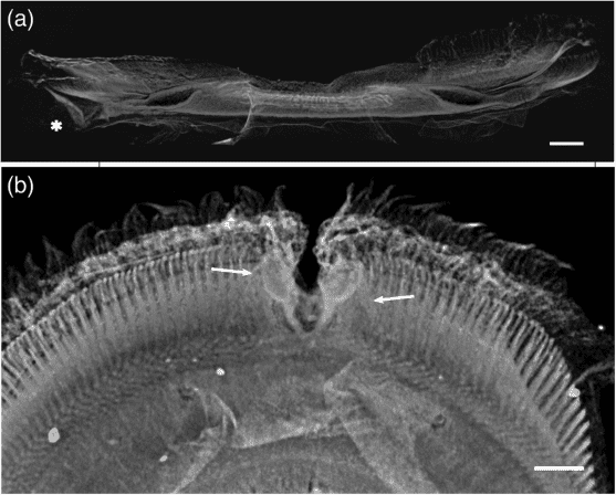
The suction discs also have a feature that hasn’t been seen elsewhere: a V-shaped notch on the rim. When this opens, the suction chamber depressurizes rapidly, enabling the larvae to lift and reposition the sucker near another patch of algae.
Video footage of moving larvae on a glass surface, taken using interference reflection microscopy, demonstrated the animals’ fine control over the V-notch. Each notch has its own pair of dedicated muscles that can be used to open it independently at various speeds.
Imaging also suggests that flaps on the V-notch are arranged in a way that creates a valve when they are closed. This prevents water from flowing into the suction chamber during attachment, helping to maintain the pressure difference.
Inspiration for engineers
Human-engineered suction cups only work well on smooth, clean surfaces, and the team hope their findings will enable them to develop more adaptable alternatives. “By understanding how the larvae’s suction organs work, we now envisage a whole host of exciting uses for engineered suction cups,” says Cambridge’s Walter Federle. “There could be medical applications, for example allowing surgeons to move around delicate tissues, or industrial applications like berry-picking machines, where suction cups could pick the fruit without crushing them.” Clingfish inspires suction cups for underwater robots
Jessica Sandoval, a materials scientist at the University of California, San Diego, who studies the suction cups of clingfish but was not involved in the present work, called the research an “exciting model” for bio-inspired suction cups. “Whether it is mimicking the microscopic microtrichia to mimicking the ‘V-notch,’ there is much to learn from the suction organ of this larvae,” she tells Physics World.
While wet environments generally make adhesion challenging, Sandoval believes that bio-inspired versions of these suction discs could make it easier to affix objects to wet, rough surfaces. Other applications are also possible. “Bioinspired suction cups that can withstand highly directional forces could be used across a wide variety of fields, from robotics to sensing,” she says. “In the field of robotics, such suction cups could be applied to manipulation or locomotion, especially in unstructured or rough terrain.”


