
An artificial intelligence (AI) algorithm can provide fully automated quantification of emphysema, offering potential as a tool for image-based diagnosis and quantification of emphysema severity, according to research published in the American Journal of Roentgenology (AJR 10.2214/AJR.19.21572).
After testing prototype AI software on over 140 patients, a multinational team of researchers found that the algorithm showed very strong correlation with traditional pulmonary function tests.
“The ability of an AI-based system to analyse high-dimensional image data and generate valuable clinical information without input from human observers should thus hold considerable potential for granular quantitative imaging approaches to provide patients with accurate diagnoses and steer treatment,” write the team, led by Andreas Fischer of the Medical University of South Carolina (MUSC) and the University Medical Centre Mannheim in Germany.
Although several visual, semiquantitative and quantitative techniques are used to assess emphysema, these methods are subject to increased interobserver and interpatient variability, according to the authors.
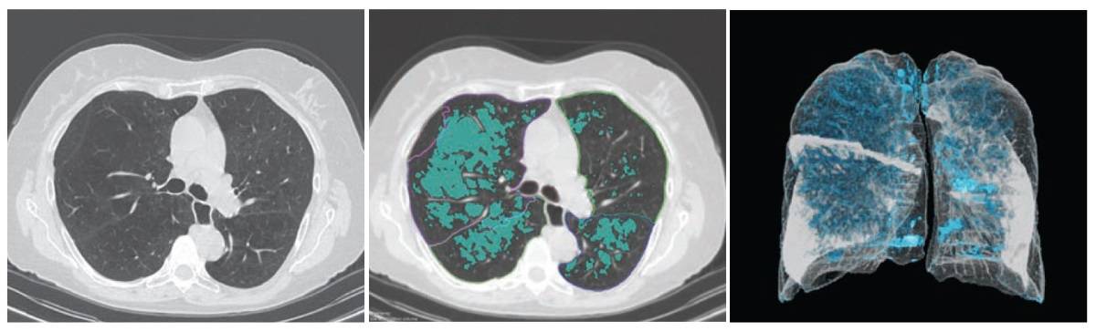
To see if AI could accurately quantify emphysema, the researchers used a deep image-to-image network – a multilayer convolutional neural network that had been trained and tested on more than 10,000 CT datasets acquired on scanners from three vendors at over 20 clinical sites in the US and Europe. Approximately 25% of these datasets were from patients with emphysema.
The algorithm was retrospectively evaluated on 141 patients who had received unenhanced chest CT and spirometry measurements within six months of each other at MUSC between August 2017 and July 2018. All CT exams had been acquired on one of three scanners from Siemens Healthineers: Somatom Definition Flash, Force or Emotion.
To determine if reconstruction methods would impact performance, the researchers applied the algorithm to two reconstruction kernels. The first method used a section thickness of 1.5 mm with a long kernel, while the second utilized a section thickness of 1.5 mm with a soft-tissue kernel. Emphysema was quantified using spatial filtering and a threshold of -950 Hounsfield units.
The patients had a mean spirometry-based Tiffeneau index (TI) of 0.57. The first reconstruction method showed a mean percentage of emphysema of 9.96 ± 11.87%, while the second method had a mean percentage of 8.04 ± 10.32%.
The algorithm correlated very strongly with the TI on both the first (Spearman correlation coefficient = -0.86) and the second (Spearman correlation coefficient = -0.85) CT reconstruction methods. Both results were statistically significant (p < 0.0001).
The results indicate that AI-based emphysema quantification meaningfully reflects clinical pulmonary physiology, according to the researchers.
“Further investigation is needed to establish quantified AI-based emphysema analysis as a potential biomarker for patients with COPD, thus improving diagnostic performance for a specific outcome and maximizing information retrieval to better understand the causes of disease,” the authors write. “Thus, AI-based pulmonary emphysema diagnostics could contribute to complementary phenotyping as part of an imaging biomarker for patients with COPD to shape the individual therapy of patients as a common diagnostic tool, in combination with pulmonary function tests.”
- This article was originally published on AuntMinnieEurope.com ©2020 by AuntMinnieEurope.com. Any copying, republication or redistribution of AuntMinnieEurope.com content is expressly prohibited without the prior written consent of AuntMinnieEurope.com.


