Women at high risk of breast cancer are more susceptible to developing cancers in the gap between routine screening exams. These so-called “interval cancers” tend to be fast-growing and account for 20% to 30% of all breast cancer cases.
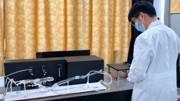
To identify aggressive cancers that develop between scheduled screenings, researchers at Massachusetts Institute of Technology (MIT) have developed a point-of-care ultrasound system that generates high-resolution 3D images of breast tissue. The 3D portable ultrasound system for real-time examination (3D PURE) supplements mammography with its ability to detect anomalies reliably in dense breast tissue (in which mammography is less sensitive).
Guided by an easy-to-use visual interface, 3D PURE enables a nurse, a physician such as a primary care provider or gynaecologist, or even the patient herself to frequently perform longitudinal breast imaging.
Second-generation design
Principal investigator Canan Dagdeviren explains that this second-generation prototype overcomes limitations of a prior system designed by the MIT team, through advances in transducer design, acoustic materials and adaptive beamforming. These developments have enabled real-time wide-angle 3D imaging in a portable form factor, with improved image resolution to accurately identify cysts, solid masses, fibroadenomas and microcalcifications.
The ultrasound system comprises a newly designed 128-element box-shaped 2D transducer array incorporating a corner-gap offset geometry, which suppresses peak crosstalk by 3.73 dB at the corner-most element. This design prevents amplifier saturation, and supports operational transmit voltages of up to 24 V. The array is integrated into a compact multilayer electronics stack comprising preamplifiers, transmit electronics and a custom chirp data acquisition system.
There’s also a conductive backing layer (a toluene-diluted polyurethane matrix with a high percentage of tungsten and zirconia filler) that forms a continuous shielding layer around the elements, improving acoustic attenuation and providing electromagnetic shielding. The compliant mechanical nature of the material enhances acoustic damping, reducing ultrasound reverberations more effectively and improving signal fidelity.
“The addition of a backing layer to the ultrasound transducer is a key advantage to the system,” explains co-lead author Md Osman Goni Nayeem. “It provides both better directionality of ultrasound waves and better bandwidth, which improves the resolution and quality of the resulting images.”
The researchers also incorporated layered aberration-correction reconstruction (LACR), an adaptive 3D beamformer, into the 3D PURE design. LACR compensates for the heterogeneous speed-of-sound in the breast, which comprises glandular and fibrous tissues plus a superficial layer of fat. They believe that this represents the first wide-angle 3D ultrasound imaging system to implement aberration-correction beamforming.
“What we are trying to do is predict the speed-of-sound properties of the tissue being imaged, and then use that to reconstruct the image more accurately. We see up to a 10% improvement for the resolution just by applying this [beamforming] technique,” says co-lead author Shrihari Viswanath in a press statement.
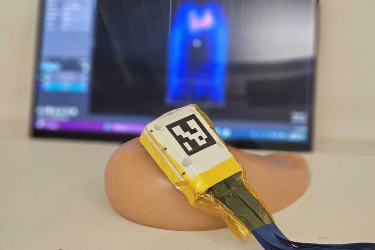
The system is designed for ease of use. A visual user interface called “Mirror my First UltraSound” (MyFUS) guides users to reliably reposition the ultrasound probe at the same anatomical location during repeated imaging and long-term monitoring. Meanwhile, a wide field-of-view minimizes the number of scans needed to cover the entire breast.
The team undertook an in vitro study comparing the performance of the 3D PURE system with that of a conventional 2D handheld ultrasound system (HHUS). Ten study participants imaged a breast-shaped tissue phantom embedded with sub-millimetre targets. Nine of the 10 participants showed improved microtarget detection efficiency with 3D PURE relative to the conventional system – identifying more of the targets using 3D PURE (79.7%, compared with 60.7% for the 2D HHUS), with a higher detection efficiency (14.02% of available targets per minute, compared with 9.58%).
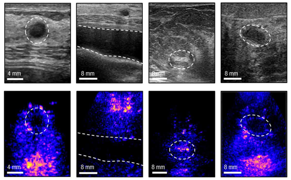
The researchers also worked with radiologists to assess a variety of breast anomalies in vivo. The radiologists verified that the 3D PURE system could accurately visualize calcifications, cysts, implants, fibrous tissues and solid masses within a large volumetric field-of-view. The researchers independently validated the system’s accuracy when imaging dense fibrous breast tissue and rib structures.
Seven volunteers with no prior experience of using an ultrasound system tested the MyFUS visual interface. After initial instruction by an ultrasound technician, they were able to repetitively position the probe at a specific location on their breasts, achieving a 94.10% mean overlap value on frontal projections, and 87.42% on side projections. This test reconfirmed that novice users could achieve reproducible longitudinal monitoring results with the aid of the visual interface.
Miniaturized ultrasound scanner could help detect breast cancer earlier

The researchers now hope to create an interface for use with mobile phones or tablets. This type of system could make breast ultrasound more accessible to patients in economically constrained countries lacking adequate breast cancer scanning and regions with shortages of trained ultrasound technicians.
“The high operator dependence of conventional ultrasound is a barrier to its use in decentralized or home-monitoring settings,” the researchers write. “The MyFUS vision interface transforms longitudinal monitoring from an expert-driven procedure to a self-guided, reproducible process that can be used by novice users. This reproducibility is vital for longitudinal monitoring in limited clinical settings, to regularly track the growth of an anomaly or the response of a tumour to therapies without visiting clinics.”
The 3D PURE system is described in Nature Communications.