Ten years ago, researchers succeeded in significantly increasing the lateral resolution of low temperature atomic force microscopy (AFM) by functionalizing the AFM tip with a single carbon monoxide (CO) molecule. This so-called bond-imaging technique was a new landmark for visualizing the atomic structure of single molecules. Unfortunately, the technique, which works by scanning the AFM tip, kept at a constant height, across a surface sample, is only suitable for flat or nearly flat molecules and not bulky, 3D ones. A team at the Justus Liebig University Giessen in Germany has now overcome this problem by exploiting the tunnelling current between the AFM tip and sample to control the tip height so that it closely follows the molecule’s topography.
Both AFM and STM (scanning tunnelling microscopy) were invented in the 1980s and both make use of a sharp tip that scans the sample surface to produce an image of it. STM measures the tunnelling current that flows between the tip and the sample while AFM exploits the force that the surface exerts on the tip. Researchers often use both techniques at the same time – STM to obtain information on a sample’s electronic structure and AFM on its atomic structure.
In bond-imaging AFM, the CO molecule at the AFM tip acts as a tiny force sensor and usually works in constant-height mode, explains Daniel Ebeling, who led this new research effort. Here, the tip that is attached to one prong of an oscillating quartz tuning fork is scanned across the surface of a molecule. This leads to small changes in the resonant frequency of the tuning fork that can be monitored. Repulsive interactions between the CO tip and the atoms of the molecules being imaged produce positive frequency shifts and bright image contrast, while attractive interactions lead to negative frequency shifts and dark contrast. In this way, an image reflecting the bond structure of the molecule can be obtained.
While the method is perfectly suited to analyse flat molecules, it falls short when imaging bulky, non-planar ones. The reason is straightforward: only the top of the 3D object can be imaged since the frequency shift is measured at a constant height and the tip is simply too far from areas underneath to collect a useful signal.
Exploiting the constant tunnelling current mode of an STM
Being able to make the AFM tip closely track a molecule’s topography would be a solution to this problem. Previous attempts to do this, however, have involved complex procedures and additional apparatus.
Ebeling and colleagues have now found a way around this stumbling block by exploiting the constant tunnelling current mode of an STM instead of operating the AFM at a constant height. Since the tunnelling current between the AFM tip and the sample surface depends on the distance between them, this means that the tip’s height can track the molecule’s topography during scanning.
“At the same time, we can achieve sub-molecular contrast by monitoring the frequency shift channel,” says Ebeling. “This simple method, which can be easily implemented on any noncontact AFM setup, allows us to directly image 3D molecular structures.”
The new technique yields the same information as the classic bond-imaging technique for flat surfaces, he adds.
Valuable add-on
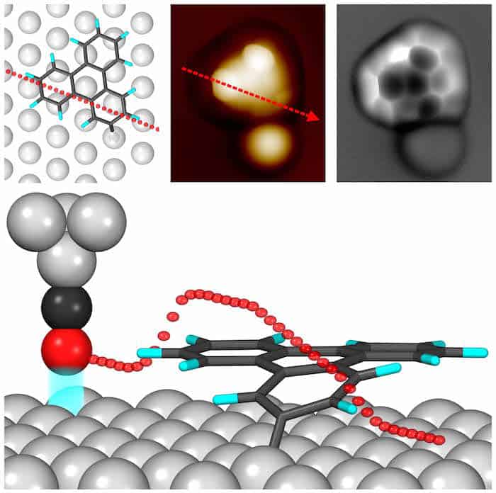
In their experiments, the researchers began by studying a flat molecule, 2-iodotriphenylene (ITP) (C18H11I) deposited on a silver (Ag) substrate. They then removed one of the iodine atoms on the molecule, which made it form a radical with a complex 3D structure.
“In this molecule, one of the carbon rings is bent towards the Ag(111) surface since the position of the radical at the corner of the molecule is strongly interacting with the Ag,” explains Ebeling. “We cannot detect this tilted carbon ring in conventional constant-height mode since it is too far away from the tip. In constant-current mode, however, it is possible to image it and even determine its tilting angle, thanks to the tip-sample feedback signal.”
The new technique represents a valuable add-on that could be widely applied in the field of surface science for studying 3D molecular systems, he tells Physics World. “Self-assembly processes, molecular recognition, and on-surface reactions of bulky compounds are some of the phenomena that could be investigated.”
Ideal for taking large overview scans
“We also show that the technique is ideal for taking large overview scans over obstacles, such as step edges, on sample surfaces without the risk of the tip crashing into them thanks to the tip-sample feedback.”
Atomic force microscope makes single-electron current meter

And that is not all. Ebeling says that the parameters of this STM feedback can be adjusted to obtain atomic-scale images of the substrate on which the molecule sits. This allows the researchers to determine molecular adsorption sites and how molecular properties are modified by the surface.
The Giessen team, reporting its present work in Physical Review Letters, has already used the technique to image aliphatic compounds such as diamondoids and for imaging nanographenes with bulky functional groups, which proves just how versatile the method is. “We will now be focusing on developing further operational modes. We are, for example, looking into whether the technique can be used with multifrequency imaging schemes, in which several eigenmodes of the tuning fork sensor are simultaneously excited,” reveals Ebeling.