
Scientists at the University of California, San Diego have used stem cells to create miniature brains that developed functional neural networks. These tiny lab-grown brains are the first observed to produce brain waves that resemble those of preterm babies, and could help shed light on the role of network activity in the developing human cortex (Cell Stem Cell 10.1016/j.stem.2019.08.002).
“The level of neural activity we are seeing is unprecedented in vitro,” says lead author Alysson Muotri. “We are one step closer to having a model that can actually generate these early stages of a sophisticated neural network.”
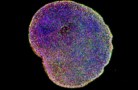
The team created cerebral organoids – a scaled-down model of the human brain, about the size of a pea – from human pluripotent stem cells. By growing them in culture medium that mimics the environment of brain development, the stem cells differentiate into different types of brain cells and self-organize into a 3D structure that resembles the developing human brain.
While researchers have successfully grown organoids with cellular structures similar to those of human brains, none of the previous models developed human-like functional neural networks. Such networks appear when neurons are mature and become interconnected, and are essential for most brain activities.
Muotri and colleagues designed an improved growth procedure, which included optimizing the culture medium formula. This allowed their organoids to become more mature than previous models. They grew hundreds of organoids for 10 months, using multi-electrode arrays to monitor their spontaneous electrical activity each week.

At about two months, the team began to detect bursts of activity from the organoids. The signals were sparse and had the same frequency, a pattern seen in highly immature human brains.
As the organoids continued to grow, they produced brain waves at different frequencies and the signals appeared more regularly. This transition suggests that the organoids had further developed their neural networks, with more functional synapses and increased connections between the neurons. The interactions between neurons contribute to signals at various frequencies.
To compare the patterns of brain waves seen from the organoids with those of human brains early in development, the team trained a machine learning algorithm with brain waves recorded from 39 premature babies between six and nine-and-a-half months old. The algorithm was able to predict how many weeks the organoids had developed in culture, suggesting that the organoids and human brain share a similar growth trajectory.
Improvements in brain organoids open new doors in neurological research
The approach provides the opportunity to investigate the emergence of network-level neurodynamics in humans. However, the researchers note that it’s unlikely these organoids have mental activities, such as consciousness.
“The organoid is still a very rudimentary model – we don’t have other brain parts and structures. So these brain waves might not have anything to do with activities in real brains,” Muotri explains. “It might be that in the future, we will get something that is really close to the signals in the human brains that control behaviours, thoughts or memory. But I don’t think we have any evidence right now to say we have any of those.”
Looking forward, the team aims to further improve the organoids and use them to understand diseases associated with neural network malfunctioning, such as autism, epilepsy and schizophrenia.


