Using a new image reconstruction technique, physicists in Germany and the US have made clear and detailed movies of nanoscale fluctuations in a magnetic material. To capture these features, a team led by Christopher Klose at the Max Born Institute, Berlin, used an advanced algorithm to identify correlations in the spatial patterns in multiple X-ray images.
Fluctuations and phase transitions are a near-universal features of matter and X-ray and electron imaging techniques can be used to observe these phenomena on the nanoscale. However, these methods have an inherent trade-off between high spatial resolution and high temporal resolution – the latter being needed to track the dynamics of fluctuations and phase transitions.
While both temporal and spatial resolution can be improved by boosting illumination, intense beams of X-rays and electrons can damage delicate features in a sample.
To overcome this limitation, Klose’s team has developed a technique called coherent correlation imaging (CCI). Their approach relies on the fact that nanoscale fluctuations are not entirely random, but instead display distinctive spatial patterns.
Many snapshots
CCI first involves taking thousands of snapshots of samples in quick succession, using relatively low levels of illumination. While these snapshots appear to be mostly indistinct from each other, the researchers found they contain enough information to categorise each image using a hierarchical clustering algorithm. This sorts the images into groups with spatial patterns that display clear correlations. By combining the images in each group, the team was able to reconstruct clear images of the patterns in samples.
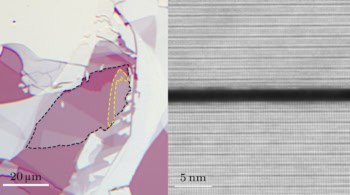
To demonstrate their approach, Klose and colleagues used CCI and X-rays to image fluctuations in a thin-film ferromagnet. This material is widely used in modern hard drives, where information is encoded into magnetic domains. These are nanoscale regions in which the magnetization can either point in one of two opposing directions. These domains are known to be highly stable at room temperature, with little information lost due to fluctuations. So far, however, researchers have not been able to confirm this stability directly by imaging the material.
X-ray microscopy sharpens up

Klose’s team used CCI to test the ferromagnet’s stability at 37 °C, which is above room temperature. Far from remaining static, the algorithm identified transitions between 30 distinct domain states in the film. By assessing the similarity between these states, the researchers also determined the order in which the transitions occurred. This allowed Klose and colleagues to construct clear, detailed movies of the fluctuations.
Through further improvements, CCI could soon enable researchers to answer fundamental questions surrounding the nature of phase transitions in advanced materials including high-temperature superconductors. Klose and colleagues now hope to extend their technique to electron microscopy – allowing them to reconstruct images on even smaller scales.
The technique is described in Nature.