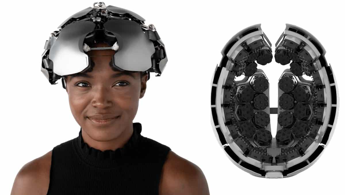
Recent years have seen huge advances in brain imaging technologies, allowing neuroscientists to explore and investigate how our brains work in more detail than ever before. To date, however, these technologies have remained in laboratory settings, with controlled experiments designed to investigate specific functions. Researchers at Kernel, a US-based neurotechnology company, hope to change this, freeing brain imaging from the laboratory and planting it in daily life. Earlier this year, Kernel researchers introduced their new device, the “Kernel Flow”, in the Journal of Biomedical Optics.
The Kernel Flow builds on the brain imaging technique of time-domain functional near-infrared spectroscopy (TD-fNIRS). fNIRS uses light in the near-infrared spectrum to measure changes in light absorption by the haemoglobin in the blood circulating in the brain. Such changes can provide information on brain function as the haemoglobin concentration changes in functioning areas of the brain because they require oxygen to power this activity. While TD-fNIRS is not a new technique, previous systems suffered from low channel numbers and slow sampling frequencies, limiting their utility in the neuroimaging field.
The researchers at Kernel designed an adjustable headset consisting of 52 modules organized onto four plates on each side of the head to provide coverage across the entire surface of the brain. Each module comprises a laser source surrounded by six hexagonally-arranged photodiode detectors that can detect more than one billion photons per second. Two lasers within the source emit light at different wavelengths (690 and 850 nm), which are directed towards the brain through the surface of the scalp.
The scattered and reflected light is then picked up by the detectors, which are placed 10 mm away from the laser source. The detected photon arrival times are recorded into histograms at a sampling rate of 200 Hz, with an overall system sampling frequency of 7.1 Hz.
The team tested the system using an optical phantom: a tank filled with a mixture of water, India ink and emulsion – with known optical properties – and a small, black PVC target placed at varying depths to mimic brain activity. This is a standard tool for characterizing the abilities of a TD-fNIRS system. The Kernel Flow performed comparably with larger benchtop systems, maintaining – or improving upon – performance whilst also being smaller and light enough to wear.
Finally, the team tested the Kernel Flow in human volunteers. Two participants took part in a neuroscientific test of the system, during which they tapped their left and right fingers in interleaved blocks with rest periods. Channels over the participants’ motor cortices showed significant haemodynamic changes during the finger-tapping tasks.

Multi-directional spectroscopy enables human neuroimaging
In addition, a channel on the forehead of one of the participants could pick out their heartbeat oscillation, an ability that is unique to this TD-fNIRS system and is enabled by its high sampling rate. These promising results have led to several follow-up studies on the application of the Kernel Flow system, including one exploring using the system to measure the effects of a psychedelic drug.
The researchers, however, acknowledge the limitations of fNIRS, and are evaluating the performance of their system on different hair and skin types, which can influence the effectiveness of optical brain imaging tools. While the Kernel Flow is not quite as commercially viable as your smart watch (yet), its introduction and the promise of commercial systems available as soon as 2024 suggests that brain function measurements may soon be as accessible as those currently used for measuring your heart rate or tracking your sleep.


