Is there something basically wrong with present-day radiology techniques? The answer would seem to be yes, on the basis of recent results from the National Synchrotron Light Source in the US.
The results show that the limited quality of conventional X-ray images can be dramatically improved by exploiting synchrotron light (D Chapman et al. 1997 Phys. Med. Biol. 42 2015). Moreover, there is no need for a high radiation dose.
Ineffective radiology has a big impact on society. Relatively high X-ray doses can overcome the limitations of conventional techniques, but they act as a deterrent for the mass screening of killer diseases like breast cancer. This is regrettable, since screening allows early detection and, in most cases, very successful therapy.
What is wrong with conventional radiology? Several things: contrast is mainly based on differences in X-ray absorption in different parts of the specimen, and these are often weak; contrast and resolution are also reduced by scattered X-rays; and finally, and more fundamentally, differences in X-ray refraction that could improve contrast are not exploited.
Now Bill Thomlinson of the Brookhaven National Laboratory and co-workers from the US, Germany and Italy have used the National Synchrotron Light Source (NSLS) at Brookhaven to develop a new approach – diffraction-enhanced imaging – that uses a highly monochromatic and collimated X-ray beam to remove some of these limitations. A double-crystal monochromator is used to filter the incident beam via Bragg scattering, and a third high-quality crystal is placed between the object being imaged and the detector to filter out X-rays that have been scattered and are therefore not travelling in the right direction. This “Bragg analyser” would, on its own, improve the image quality, but the NSLS approach goes beyond this simple improvement and ingeniously exploits X-ray refraction to produce even better images.
Refraction by the specimen slightly changes the X-ray direction. However, the refracted X-rays are not rejected by the Bragg analyser because it has a finite angular acceptance. Rather, the angular response or “rocking curve” of the analyser creates contrast between regions of the sample with different refractive indices. The rocking curve is a bell-shaped plot of transmission against angle of incidence and has a maximum at an angle defined by the X-ray wavelength and the lattice spacing in the crystal.
In an unprocessed image, conventional absorption-contrast effects are mixed with the contrast due to refraction. Image processing can, however, separate these effects. To do this, two different images are taken when the Bragg analyser is rotated to the two points of maximum slope on the rocking curve: simple algorithms are then applied pixel-by-pixel to the images. A pair of raw images can thus yield two processed radiographs of superior quality, each based on a different contrast factor. The enhancement is quite spectacular (see right image).
The contrast in one of the processed images is due to absorption, or more precisely “apparent absorption”, a combination of absorption and the intensity loss caused by diffraction as the beam passes through the specimen. The exploitation of this intensity loss, known as extinction, can significantly increase the information obtained from the radiograph.
The other image-processed radiograph – the refraction-contrast image – shows the boundaries between regions with different refractive indices particularly clearly. The processing creates a pseudo-shadowing at the boundaries and an intriguing three-dimensional appearance. The two processed radiographs provide complementary information, mutually enhancing their effectiveness.
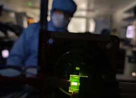
Diffraction-enhanced imaging is not the only way to produce superior radiographs with synchrotron light. A novel technique called “phase-contrast imaging” has been developed at the European Synchrotron Radiation Facility in Grenoble, France (A Snigirev et al. 1996 Nucl. Instrum. Meth. A370 634). This approach was recently implemented on the ELETTRA synchrotron at Trieste in Italy by Edoardo Castelli, Ludovico Dalla Palma, Giuliana Tromba and collaborators at a radiation dose compatible with medical radiology (see left image). Compared with a conventional radiograph, a phase-contrast image exhibits very sharp and highly visible boundaries. This can simplify the early detection of microstructures in cancer screening, defect analysis in technological components and many other applications.
There is still some uncertainty about what causes the contrast enhancement, but many authors agree that the temporal and spatial coherence of the X-ray beam are the key factors. Specifically, it is thought that diffraction at the edge between regions with different refractive indices produces sharp diffraction fringes that highlight the boundaries. However, in addition to a coherent X-ray beam, this mechanism also requires a detector with sufficient spatial resolution to reveal the fringes.
Temporal coherence of the X-ray beam can be achieved by brute force, by simply making the beam monochromatic. In fact, virtually any monochromator can provide the necessary temporal coherence. Lateral or spatial coherence is both a novel and exciting factor in X-ray science, and has been made possible by the new synchrotron sources. Indeed, the same geometric characteristics that lead to high brightness – small source size and small angular divergence – also lead to spatial coherence. The source sizes (which are related to the lateral dimensions of the electron beam producing the synchrotron radiation) that are found in most of the new synchrotron sources are much less than 100-200 µm, which leads to adequate spatial coherence for phase-contrast imaging.
The coherence-based explanation and even the term “phase contrast” are questioned by some authors. For practical applications, however, a full explanation of the contrast mechanism is largely irrelevant: Röntgen took radiographs of his wife’s hands without knowing anything about atomic structure or X-ray absorption! What really matters is that a significant increase in contrast can be achieved with synchrotron light, and this has been solidly demonstrated in experiments.
Can synchrotron sources actually be used for routine medical radiology? The barriers are more psychological than real, even from the financial point of view: special synchrotron facilities entirely dedicated to medical facilities are certainly conceivable. Indeed, similar doubts were probably raised one century ago about Röntgen’s apparatus. One can hope, therefore, that in the future, radiology will not be based on a high dose of radiation, but on a high dose of creativity.