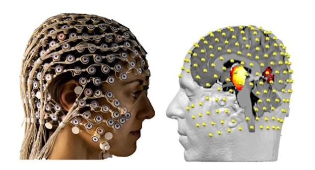
Subcortical brain structures such as the thalamus and nucleus accumbens play a critical role in controlling motor, emotional and associative activity. Dysfunction in communication between these structures and with the cortex can lead to serious diseases, including Tourette syndrome, obsessive-compulsive disorders (OCD) and Parkinson’s disease. However, it is extremely difficult to study these structures, which are located deep inside the brain, and it’s not precisely clear how they work.
Current treatments for these diseases are based on deep brain stimulation (DBS), an invasive approach that uses electrodes implanted into the centre of the brain. DBS has been shown to be effective in Parkinson’s, but doesn’t work so well for OCD and Tourette’s. Understanding how subcortical brain zones function and communicate could help improve treatments, but existing approaches for measuring activity in deep brain areas are also highly invasive.
As such, researchers from the University of Geneva (UNIGE) and Cologne University investigated whether electroencephalography (EEG) — a non-invasive method that records the brain’s electrical activity via electrodes placed on the scalp — could measure deep brain activity from outside the skull. They demonstrated, for the first time, that EEG combined with mathematical algorithms can record signals usually only seen by implanted electrodes (Nature Communications 10.1038/s41467-019-08725-w).
Simultaneous measurement
To determine whether activity in deep brain structures can be sensed using EEG, the researchers simultaneously recorded subcortical activity using the implanted electrodes and high-density (256-channel) EEG scalp electrodes. They examined two patients suffering from Tourette’s and two OCD patients, who had DBS electrodes implanted in the thalamus and nucleus accumbens, respectively.
The results from the two measurement techniques correlated perfectly. “The mathematical algorithms that we developed meant we could accurately interpret the data provided by the EEG and ascertain where the brain activity was coming from,” explains first author Martin Seeber.
“In obtaining highly similar signals as with the implants, we finally proved that surface EEG can be used to see what is happening in the deepest part of the brain without having to go into it directly,” adds senior author Christoph Michel.
The authors note that previous work using simulations and source reconstruction provided indirect evidence for the detectability of subcortical sources in EEG recordings. “In this work, we can confirm this finding with direct evidence from intracranial recordings providing the ground truth of subcortical signals, in combination with non-invasive EEG source reconstruction locating these signals in close proximity to the actual recording sites,” they write.
With this confirmation, the researchers can now try to understand how subcortical structures communicate with each other and with the cortex, hopefully leading to increased understanding of the causes of diseases such as Tourette’s and OCD. The team is also keen to use the technique to improve existing treatment methods, based on rebalancing network interactions using a very slight electric shock. They would also like to address other disorders, such as obesity, addiction or Alzheimer’s disease.
“Lastly, we hope that in time we’ll be able to stimulate the deep brain areas from the surface via an electromagnetic treatment, doing away with electrode implants in the brain once and for all,” says Michel.


