
Fatal and non-fatal injuries caused by landmines and improvised explosive devices (IEDs) are appallingly common, afflicting as many as 20,000 people per year in current and historical conflict zones. Those who survive such blasts typically suffer blast-mediated amputations caused by the high-pressure shock wave and other explosive effects.
In addition to the immediate damage that it causes to the limb, the shock wave also triggers a long-lasting condition that emerges in the weeks and months after the blast. This condition – heterotopic ossification (HO) – is the anomalous formation of bone in soft tissues such as muscles and ligaments, usually in the parts of the limb closest to the site of the initial injury.
“HO is a major problem,” explains David Sory from Imperial College London. “It poses one of the most significant clinical problems to casualties suffering from blast-mediated amputation, as this pathology has a direct impact on rehabilitation and return to functional mobility.”
Despite HO’s prevalence among blast survivors, the details of how mechanical loading of tissues induces the condition are still not known. This is largely because experiments involving living cells have not yet encompassed the extreme stress regime associated with blast trauma. Sory and colleagues sought to fill that gap. By bringing together expertise from the fields of shock physics and stem-cell biology, the team developed a new experimental setup to study the cellular effects of extreme mechanical loading.
The researchers built three separate loading platforms that can subject in vitro cell and tissue samples to a range of stresses, from those caused by everyday activities at the lower end, to the kind of forces caused by landmines and other explosive devices at the higher end. In their first experiments using the new setup, which took place in the Centre for Blast Injury studies at Imperial College, they found that gene activity denoting HO has a complex relationship with loading parameters.
The team adapted existing experimental apparatus so that they could accommodate the variety of cellular samples commonly used in in vitro investigations. These samples need to be maintained in a sterile and biocompatible environment for the duration of the experiment, and to be retrieved intact for study afterwards.
They fulfilled these requirements by encapsulating the samples in hermetically sealed pressure chambers formed from polydimethylsiloxane (PDMS). By controlling the ratio of its components, the researchers tuned the polymer’s acoustic impedance to match that of native tissues, enabling pressure waves to pass efficiently from the apparatus to the cells within the sample.
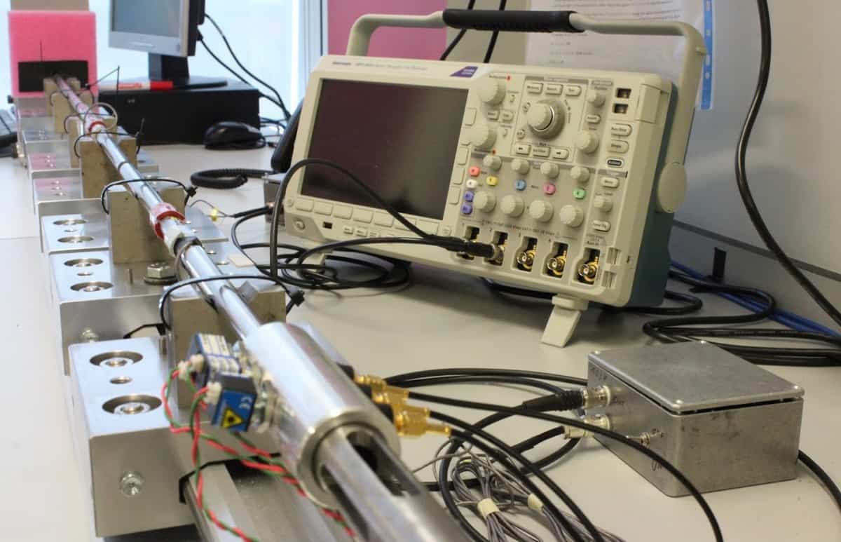
To span a wide range of strains and strain rates, the researchers integrated the pressure chambers into three separate experimental platforms. The physiological strain-rate regime – representing stresses like those induced by walking, running and jumping – was handled by a modified universal testing machine. A drop-weight rig covered the intermediate regime, and a split-Hopkinson pressure bar provided extreme loading rates like those experienced by tissues near to an exploding landmine or IED.
The team’s first test of their new setup involved mesenchymal stromal cells (MSCs) obtained from rat periosteum, the thin tissue membrane covering the outer surfaces of bones. In some experiments, the researchers suspended these cells in fluid; in others, the cells were held within 3D hydrogel scaffolds. Twenty-four hours after subjecting the samples to a range of strains and strain rates, they measured the expression of a transcription factor called Runx2, which indicates that the cells are primed to differentiate into bone tissue — as happens in HO.
While Runx2 expression was related to the mechanical loading – only intermediate and high strain rates produced an increase, for example – the pattern was not straightforward. Rather, the strain rate, peak strain, strain duration and the nature of the sample (fluid suspension or 3D scaffold) all combined to contribute to priming the cells in a complex way.
In future experiments, Sory and colleagues hope to explore the cellular mechanics of HO more thoroughly using human MSCs in samples that better represent in vivo tissues. Although their ultimate aim is to develop therapies that prevent the onset of HO in blast victims, there could be circumstances where it would be useful to trigger the condition deliberately.
“In fact,” says Sory, “understanding the mechanisms of HO formation could help develop novel therapeutic strategies for bone diseases and to repair bone loss.”
Full details of the research are reported in Physical Biology.


