Diagnosing Alzheimer’s disease remains a challenge, with no practical tools available for early detection or large-scale screening. Instead, the disease is often diagnosed using memory tests or by observing behavioural changes. But by the time symptoms have appeared, the disease is advanced. What’s needed is a rapid, non-invasive and inexpensive way to detect Alzheimer’s at the earliest stages.
With this aim, researchers from the Duke Eye Center have demonstrated that optical coherence tomography (OCT) angiography can detect signs of Alzheimer’s disease. In a study of more than 200 people, the non-invasive imaging technique showed that blood vessels in the retina are altered in patients with Alzheimer’s. OCT angiography could also distinguish between people with Alzheimer’s disease and those with mild cognitive impairment (Ophthalmology Retina 10.1016/j.oret.2019.02.002).
OCT angiography enables visualization of the smallest blood vessels in the back of the eye, which are thinner than the width of a human hair. The scan could reveal changes in these tiny capillaries before they can be seen on an MR brain scan or cerebral angiogram, which only highlight larger blood vessels. Because the retina shares many similarities with the brain, researchers believe that deterioration in retinal microvasculature may mirror changes in blood vessels in the brain, thereby offering a window into the disease process.
“We know that there are changes that occur in the brain in the small blood vessels in people with Alzheimer’s disease,” explains lead author Dilraj Grewal. “Because the retina is an extension of the brain, we wanted to investigate whether these changes could be detected in the retina using a new technology that is less invasive and easy to obtain.”
“We’re measuring blood vessels that can’t be seen during a regular eye exam,” adds senior author Sharon Fekrat, “and we’re doing that with relatively new non-invasive technology that takes high-resolution images of very small blood vessels within the retina in just a few minutes.”
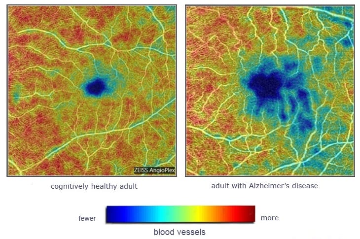
Grewal, Fekrat and colleagues used OCT angiography to image the eyes of 39 Alzheimer’s patients, 37 people with mild cognitive impairment (often a precursor to Alzheimer’s disease) and 133 cognitively healthy people. In the healthy group, the researchers saw that microscopic blood vessels form a dense web at the back of the eye inside the retina. In Alzheimer’s patients, that web was less dense, with a loss of these small retinal blood vessels.
They also found that a specific layer of the retina was thinner in the Alzheimer’s group than in people with mild cognitive impairment and healthy controls. The differences in density were statistically significant after the researchers controlled for factors including age and sex.
Portable imager provides insight into eye and brain diseases

“Early diagnosis of Alzheimer’s disease is a huge unmet need,” says Fekrat. “It’s not possible for current techniques like a brain scan or lumbar puncture to screen the number of patients with this disease. It is possible that these changes in blood vessel density in the retina may mirror what’s going on in the tiny blood vessels in the brain. Our work is not done. If we can detect these blood vessel changes in the retina before any changes in cognition, that would be a game changer.”
The ultimate goal, says Fekrat, is to use OCT angiography to detect Alzheimer’s disease early and to monitor these changes over time in participants of clinical trials studying new Alzheimer’s treatments.


