Researchers in Germany and the UK have developed a real-time imaging technique to capture neuronal dynamics in deep-brain layers of living mice, achieving a resolution of around one micron. The high-speed fibre-based fluorescent imaging probe is capable of recording subcellular neuronal structures in the most minimally invasive manner reported to date (Light: Science and Applications 10.1038/s41377-018-0094-x).
Advances in spatial light modulation technologies have led to promising progress in biomedical research. An encouraging example of this is the use of multimode fibres (MMFs), which work as ultranarrow endoscopic probes that allow high-resolution imaging. MMFs also overcome the limitation on the size of optical elements used deep inside living tissues, leaving no structural and functional impact.
Currently, the deep layers of the visual cortex and the hippocampus are fairly inaccessible due to lying deep within the brain. While current massive endoscopes with hundreds of optical fibres are too invasive to penetrate deep-brain regions composed of very tiny structures, non-invasive imaging methods like MRI cannot resolve the tiny neurons within sensitive brain areas. Therefore, designing a compact and carefully optimized imaging system for time-lapse observation and study of neuronal connectivity in vivo could enable a new level of potential biomedical investigations.
Multimode fibre-based imaging system
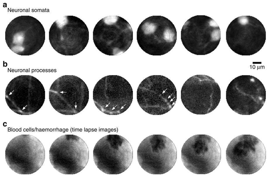
The researchers — from the Leibniz Institute of Photonic Technology (Leibniz IPTH) and the University of Edinburgh (Centre for Discovery Brain Sciences), led by Tomáš Čižmár and Nathalie Rochefort — took advantage of high-tech holographic methods to achieve single fibre-based imaging of neuronal subcellular processes over several hours. They achieved a resolution of about 1.18 µm across a 50 µm imaging field-of-view, at 3.5 frames/s.

The 2 cm long fibre was post-processed into a flat-cone to minimize destructive tissue compression as the fibre penetrated to more than 2 mm below the brain’s surface. To confirm the extent of damage after fibre penetration, the researchers looked at a post-mortem section of a perfused brain and saw minimal tissue damage. In addition, single fibre-based imaging not only shortens the post-operative recovery period but also eliminates the need to implant optical imaging elements such as a graded index lens.
“We are very excited to see our technology making its first steps towards practical applications in neuroscience,” says lead author Sergey Turtaev from Leibniz IPTH. Furthermore, Rochefort, the co-supervisor of the project, believes that one of the future applications of this new minimally invasive approach would be to observe neuronal activities in deep-brain structures in behaving animals. This way, neuroscientists will hopefully be able to investigate the many remaining knowledge gaps regarding memory formation and sensory perception, for instance.
Despite the many optimizations that were performed on the imaging system, a considerable level of background was still visible in the obtained images. The origin of this background is the out-of-focus light at the time of sample excitation. “Eliminating out-of-focus light by means of confocal rejection of incoherent fluorescent signals returning from the MMF is currently not possible without prohibitive power losses. Therefore, future work will focus on the development of computational post-processing algorithms to further enhance the imaging quality,” the authors conclude.


