Asylum Research was built on its founder’s zeal for biophysics — but now its atomic force microscopes are pointing the way towards denser computer memories, and one even has appeared in one of the world’s most popular television shows. Hamish Johnston speaks to co-founder Roger Proksch.

When physicists Roger Proksch and Jason Cleveland founded Asylum Research in 1999, little did they know that nine years later one of their atomic force microscopes (AFMs) would be featured in one of the most successful US television programmes of all time — Crime Scene Investigation (CSI).
The company’s Molecular Force Probe-3D was used to produce a high-resolution image of a drug capsule found during a murder investigation in the episode “Rock and a hard place” of CSI: Miami. While the real-life applications of the firm’s systems are perhaps not as sexy as an appearance on CSI, Proksch told physicsworld.com that he continues to be surprised by the growing number of ways that researchers are using Asylum’s equipment.
Before founding the company, Proksch and Cleveland worked at US-based Veeco. However, the firm was becoming increasingly focused on making metrology equipment (including AFMs) for the semiconductor industry — something that didn’t appeal to the two physicists.
Dabbled in biophysics
“We were interested in making instruments for biophysics at the time,” recalls Proksch. Although the pair both did PhDs in the use of AFMs to study magnetic materials (Cleveland at the University of California at Santa Barbara and Proksch at the University of Minnesota) they had a growing interest in biophysics, having both “dabbled” in the field as postdocs. So they joined forces with lawyer Dick Clark to found Asylum Research, which has its headquarters in Santa Barbara, California. They now have about 60 employees, including many with PhDs in physics.
The firm’s first instrument was not an AFM, but rather a system that allowed biophysicists to measure the forces that hold large molecules together. This is done by attaching one end of the molecule to a substrate and the other to the tip of a tiny cantilever. The cantilever is then used to carefully pull the molecule apart, while the forces required to do so are determined by monitoring minute deflections of the cantilever.
This technique is called single-molecule force spectroscopy and can be used to study the structural properties of a wide range of large molecules such as DNA or polymers. Smaller molecules can also sometimes be studied by engineering them into larger molecules — using the larger molecules as “handles” to pull on the smaller molecules.
In 1999 the technique was only a few years old, recalls Proksch: “For us it was a nice niche, it allowed us to do something that was related to our experience but new enough that there was no competition”. The company ended up selling about 70 force spectrometers, the vast majority going to university biophysics research labs. Asylum had hoped that its instrument would become a diagnostic tool in the pharmaceutical industry. “But that just hasn’t happened and I’m not sure it will ever happen for this technology”, says Proksch.
Customer requests
By about 2002, sales to universities were beginning to slow down and Asylum saw its next opportunity in a request that it was getting from many of its customers — the ability to precisely scan the tip in the x–y direction, as well as up and down. Although the height of cantilever above the substrate could be positioned with nanometre accuracy, the instrument had relatively low resolution in the x–y plane. This meant that the user couldn’t know for certain exactly where on the molecule they where pushing or pulling.
“Our customers were asking us to make a system that could create an image of the molecule before you started poking at it — and that is basically an AFM”, says Proksch.
Fortunately, Asylum could use some of the same technology it had developed for the accurate positioning of the height of the cantilever to scan it in the x–y direction across the sample. This led to the development of the firm’s current “flagship” instrument, the MFP-3D. “One of the strengths of this instrument is that you can take these wonderful images of the sample and then you can go back and examine the other properties at specific locations”, says Proksch.
Proksch puts much of the firm’s success down to its use of linear variable differential transformer (LVDT) position sensors for use in its instruments. An LVDT comprises a primary coil with a current flowing through it to create a magnetic field. Some of the magnetic flux passes through two secondary coils, inducing currents in these coils. One contact of the position sensor is connected to the primary coil and the other contact to the two secondary coils. If the distance between the contacts changes, so does the relative positions of the coils and this change can be determined by measuring and comparing the induced currents in the secondary coils.
This is unlike its competitors, which use sensors based on capacitive plates to determine the position of the cantilever, if they use a sensor at all. The main difference between Asylum’s LVDTs — which use mutual inductance between magnetic coils — and low-noise capacitive sensors is that the latter require plates that are very large and in a perfect parallel planar configuration, which is very difficult to achieve, according to Proksch. “Magnetic coils are much more forgiving in terms of geometry”, he explains.
Low-noise amplifiers
Most LVDTs use a primary coil with a ferromagnetic core, which increases its magnetic field thereby boosting the sensitivity of the sensor. However, the sensitivity of this structure is limited by “Barkhausen noise” in the core, which results in sudden random changes in the magnetic field. To get around this problem, Asylum does not use a core in its patented LVDTs, but rather boosts the sensitivity of the secondary coils by using the latest in extremely low noise amplifier technology.
As a result, the sensors have noise level of about 0.1 nm noise in a 1 kHz bandwidth, which means that the instrument can make one position measurement per millisecond with an uncertainty of 0.1 nm.
While capacitive sensors claim to offer similar noise levels, Proksch says that they are much more difficult to integrate with an AFM. LVDTs, he says, can be used in a number of different geometries, giving Asylum considerable flexibility in designing their instruments — something that can be used to improve their overall performance. “We are the only AFM maker that I know of that takes this magnetic approach”, says Proksch, adding “I believe we are the only company that makes its own position sensors and this gives us the expertise to put our sensors just about anywhere”.
Growth in materials science
By creating instruments that scan in 3D, the firm has expanded its customer base beyond biophysics — which today is about 40% of its business — and has enjoyed significant growth in materials science.
The study of the piezoelectric properties of materials is one area where Asylum has seen significant growth. Piezoelectric materials generate an electric potential when squeezed or stretched — and such materials squeeze or stretch themselves when an electric potential is applied.
Although physicists have long known that many common materials are piezoelectric — including bone, wood, and many minerals — it had been very difficult to make detailed studies of the relationships between the microscopic structure of a material and its piezoelectric properties. This is particularly important to researchers trying to develop ferroelectric memories, in which data are stored in bits defined by the electric polarization of tiny domains in a ferroelectric material.
Today’s ferroelectric memories have a relatively low density and to improve this, researchers must be able to reduce the size of ferroelectric domains in the material.
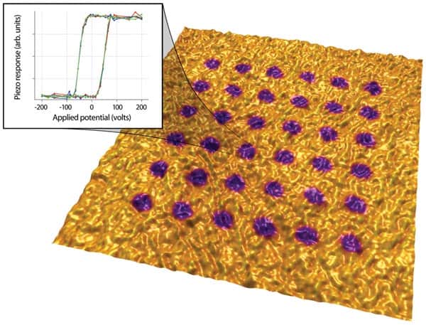
An AFM can be used to study the piezoelectric properties of a material by bringing the tip in contact with the surface, applying an electric potential between the tip and the material, and then measuring any movement in the material by watching for a deflection in the position of the tip. This technique is called piezo force microscopy or PFM.
According to Proksch, the MFP-3D can be used in piezoelectric scanning mode to image ferroelectric domains at resolutions down to several nanometres and garner additional structural information about ferroelectrics that could lead to denser ferroelectric memories.
Understanding ‘bioelectricity’
Beyond the development of ferroelectric memories, Proksch believes that the piezoelectric response of biological materials is a new and exciting area of growth for Asylum’s AFM. The piezoelectric properties of bone, for example, could be related to healing processes, whereby structural changes in damaged bone send out electrical signals telling surrounding cells to increase bone mass. As a result, such studies could lead to a better understanding of the role of “bioelectricity” in living organisms.
Proksch told physicsworld.com that the company is also currently working on reducing the time that it takes to obtain an image, which is typically about five minutes. This can be done by increasing the oscillation frequency of the tip, which in practical terms means making the cantilever smaller. While this is straightforward in principle, in practical terms it means that the rest of the hardware and software on the instrument also operate much faster — which is a significant challenge that the firm hopes to meet.


