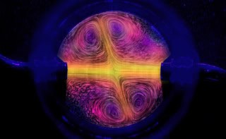
Fractal patterns that arise when healthy human cells turn cancerous have been observed for the first time by scientists in the US. Using an atomic force microscope (AFM), Igor Sokolov and colleagues at Tufts University and Clarkson University saw the patterns while studying the surfaces of cervical epithelial cells at nanometre resolution. The work could give us a better understanding of how the surface of cells affects the progression of some cancers, which could in turn lead to new strategies for fighting the disease.
Although the origin of many cancers is still a mystery, some scientists believe that these diseases are linked to complex processes in living cells becoming unbalanced, which could lead to chaotic behaviour. Indeed, signs of chaos have already been seen in biochemical and physical studies of cancerous tissue – with the structure of some cancerous tissues, for example, having fractal properties associated with chaotic systems.
Fractal patterns had, however, never been seen before on the surfaces of single cancer cells. The new observation could be significant because scientists already know that the surface of a cancer cell plays an important role in “metastasis”. This is the process whereby cancer cells manage to leave a primary tumour – often forcing their way through healthy tissue – and travel to other parts of the body to create secondary tumours.
Immortal cells
The new study was carried out using cells cultured in the laboratory. Three types of human cervical epithelial cells were studied: normal cells taken from healthy women; malignant cancer cells taken from cancer patients; and “immortal” pre-cancerous cells that were created by treating some of the healthy cells with a human papilloma virus genome. The cells were then freeze-dried so that they could be studied with an AFM.
The researchers mapped the structural features of the surfaces of the cells at a resolution of less than about 20 nm per image pixel. In particular, the AFM measured the “stickiness” between the instrument’s tiny probe and the cellular surface. The images were then processed using a Fourier transform to identify any repeating patterns. The team then analysed this information for signs of patterns that repeat on a number of different length scales – a hallmark of a fractal pattern.
The team found that the surfaces of both healthy cells and cancer cells did not have fractal patterns, whereas such patterns were seen on the pre-cancerous cells. This finding was unexpected. “Despite previous expectations that fractal patterns are associated with cancer cells,” says Sokolov, “we found that fractal geometry only occurs at a limited period of development when immortal cells become cancerous.”
Surface transformation
According to Sokolov, the team also discovered that cells deviate more from fractal behaviour when they further progress towards cancer, while normal cells do not have fractal patterns. This could mean that the fractal pre-cancerous phase plays a role in transforming the surface of a healthy cell to that of a cancer cell.
Sokolov and colleagues hope that their discovery could help to identify “weak points” in the transition from healthy to cancerous cells that could be targeted to stop the development of cancer. Such a transition could involve instabilities in biological processes that occur in the cell and lead to chaotic behaviour at the surface. If these instabilities could be prevented from emerging, then the progression to cancer could be halted.
“We need to further our understanding as to how important the cell surface is in the development of cancer,” concludes Sokolov.
The research is described in the New Journal of Physics.
- There is more about how AFMs are being used to study cancer cells in the July 2013 special issue of Physics World on the physics of cancer.