
A new study has shown that the representation of hand movements in the brain continues to exist in above-elbow amputees, years after amputation. Researchers used machine learning (a linear classifier) to “decode” the brain activity related to attempted hand gestures performed by the phantom limb, compared with the amputees’ intact hand, as well as gestures executed by non-amputee controls. This work lends support to the use of decoded brain activity patterns in the development of brain-computer interfaces (Brain 10.1093/brain/awx274).
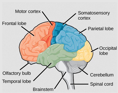
The cortical representation of touch and movement are encoded in somatosensory and motor brain regions, respectively; together, they are known as the sensorimotor cortex. Previous studies using functional MRI (fMRI) have shown activity in said cortex among amputees, indicating that, following loss of nerve supply (denervation) to the limb, the responsible brain regions are still operative.
However, it is unclear how detailed and specific the activity is, and whether complex “attempted” movements by the phantom limb can be discriminated. Here, researchers from the Brain Center Rudolf Magnus at UMC Utrecht decode brain activity corresponding to six attempted hand gestures during a fMRI scan.
Decoding brain activity
A typical fMRI analysis comprises inputting voxel-wise data (each 3D “pixel” in the brain will have a time-series of activity) into a statistical model, then comparing the outputted model weights to every other voxel in the brain. If the test statistic for this weight is significantly above chance, then that voxel is said to be “active”. Unfortunately, this methodology is unable to look at patterns of activity derived from multiple voxels.
Multi-voxel pattern analysis (MVPA) is one way to overcome this. In the simplest case, a machine learning algorithm, known as a classifier, is trained on a subset of the data and is then used to classify the remaining data into provided categories, such as faces and houses, for example.
Phantom movements are preserved
In this study, the researchers decoded the fMRI BOLD signal changes (an indirect measure of neural activity) resulting from six different attempted gestures, in four sub-regions within the sensorimotor cortex. The classification scores were well above what could be expected if (obtained at random or) obtained by chance. This suggests that there is information about the hand movements contained in these sub-regions, even for “movements” of the phantom hand in amputees.
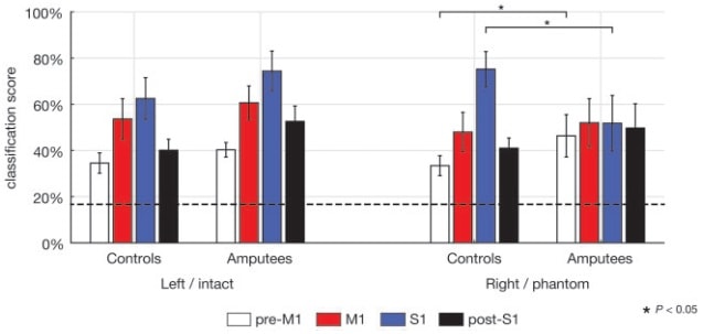
When comparing activity from the amputees’ phantom hand with that from the controls’ right hand, the team saw no difference in classification score within the primary motor cortex (M1), indicating the preservation of hand movement in amputees in M1. The high decodability of the primary somatosensory cortex (S1) – albeit lower in amputees than in controls – supports the idea that hand representations are also preserved in S1 after denervation, even after several years.
Future work investigating the topographical organization of these phantom movements, and how the sensorimotor cortex compares to other cortices that display this trait, for example the retinotopic visual cortex, would be extremely interesting. This study provides support for the development of brain-computer interfaces, which could use the decoded data, possibly using “neuro-feedback” techniques, to pave the way towards the integration of high-tech prosthetics into our human “wet-ware”.


