A diamond-based sensor that maps out the subtle electrical currents inside the heart has been developed by researchers in Japan. Led by Takayuki Iwasaki at the Tokyo Institute of Technology, the team based its device on the fluorescence of nitrogen-vacancy (NV) centres in diamonds. They used their sensor to measure the magnetic fields created by electrical currents travelling in the hearts of living rats and the researchers say that the device’s 5.1 mm resolution is unprecedented.
Some heart diseases including tachycardia and fibrillation are caused by imperfections in how electrical currents are conveyed through the heart. To diagnose these conditions, cardiologists use magnetocardiography (MCG): a contactless technique the remotely measures the magnetic fields produced by electricl currents in the heart.
The resolution of MCG is limited by factors including sensor size and operating temperature. For example, superconductor-based sensors are very good at detecting small magnetic fields, but they must be kept at very low temperatures. As a result, these sensors must be kept some distance away from the heart and therefore cannot resolve currents on the millimetre scale. This means that they cannot fully resolve the intricate rotational waves produced by ventricular arrhythmias.
Atomic-scale defects
To create a higher-resolution sensor, Iwasaki’s team used nitrogen vacancy (NV) centres – which are atomic-scale defects in diamond. In an NV centre, a pair of adjacent carbon atoms in the diamond lattice is replaced by a nitrogen atom and an empty space. An NV centre is essentially an isolated quantum spin that is very sensitive to an external magnetic field. What is more, it emits fluorescent light in a way that is dependent upon the intensity and direction of the field. These properties can be combined to create a magnetic sensor with an optical readout.
Quantum sensor could detect SARS-CoV-2
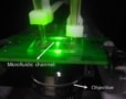
Iwasaki and colleagues created a sensor from a diamond chip with a high density of NV centres. Operating at room temperature, they positioned the sensor just a few millimetres from the hearts of live rats. The NV centres were illuminated with a green laser and a photodiode was used to capture the emitted fluorescent light. Iwasaki’s team also developed a mathematical model to translate the fluorescence measurements into the corresponding magnetic fields. This allowed them to produce detailed 2D images of electrical activity in the hearts, achieving a resolution of 5.1 mm. The researchers hope that their sensors could make it far easier for cardiologists to study the origin and progression of many different types of heart condition in patients – potentially leading to new methods for diagnosing and treating these diseases. With further improvements, the sensor could also be used to detect even more subtle electric currents produced in other parts of the body.
The research is described in Communications Physics.