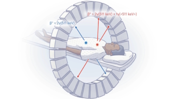
A PET radiotracer that can detect deep vein thrombosis (DVT) in the legs and clots that have travelled to the lungs has been chosen as the “Image of the Year” at the Society of Nuclear Medicine and Molecular Imaging (SNMMI) 2026 annual meeting. Developed by Sangwon Han and colleagues at the Asan Medical Center, University of Ulsan College of Medicine, in Korea, the novel tracer enables whole‑body imaging of blood clots (thrombi) in the legs and lungs in a single scan.
A DVT is a blood clot that forms in a deep vein, usually in the legs. It’s a common condition, with an incidence of roughly half that of all cancers, and it can lead to serious complications. Clots can break off and travel to the lungs, which could cause a potentially life‑threatening pulmonary embolism (a blockage in the artery supplying blood to the lungs). Early detection of DVT is therefore critical for determining the most appropriate treatment for each patient.
Currently, the standard imaging method for diagnosing DVT is venous ultrasonography (VUS). But while this works well for detecting clots in the thigh-to-knee region, whole-leg VUS requires skilled operators and advanced machines, takes longer, and has lower diagnostic sensitivity in the calf. In addition, conventional imaging techniques such as VUS and CT rely on indirect structural changes rather than directly visualizing the clot.
Aiming to enable faster and more efficient DVT diagnosis, Han and his research team are studying fluorinated GP1 (18F-GP1) a novel thrombus-targeted PET tracer. The tracer selectively binds to specific receptors on activated platelets (the cell fragments that cause blood to clots), allowing direct visualization of active thrombus formation.
“In our Phase 1 study, 18F-GP1 PET/CT showed 100% detection rate in 20 patients with confirmed DVT or pulmonary embolism,” Han told the SNMMI delegates. “But that study was limited by its small sample size and an absence of negative groups so specificity could not be assessed at that time.”
So in this latest work, Han and his team performed a phase 2, non-randomized study investigating the ability of 18F-GP1 PET/CT to identify acute lower-extremity DVT in 46 symptomatic patients. This included 22 patients with proximal DVT and 24 with none or distal DVT, as diagnosed using VUS.
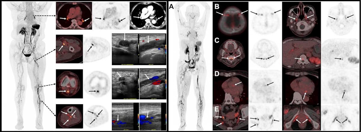
The researchers acquired chest-to-feet PET/CT scans approximately 2 h after intravenous administration of 250 MBq of the radiotracer. The images were assessed by three blinded nuclear medicine physicians from different institutions, who assigned focal 18F-GP1 uptake higher than background activity as positive for thrombosis. They classified proximal DVT as clots involving the iliac (pelvic), femoral (thigh) and popliteal veins (behind the knee), and distal DVT as clots confined to the calf veins.
“Our primary objective was to assess the sensitivity and specificity of qualitive 18F-GP1 PET/CT interpretation for proximal DVT,” Han explained. “Secondary objectives included assessing the agreement between PET/CT and VUS for distal DVT, inter-reader reproducibility, exploring the detection of pulmonary embolism and assessing safety.”
When evaluated against VUS as a reference standard, 18F-GP1 PET/CT exhibited high diagnostic accuracy for detecting clots, demonstrating a sensitivity of 95% and a specificity of 92% for proximal DVT. “For distal DVT, both positive and negative agreement between PET/CT and VUS were strong,” added Han. “Inter-reader agreement was also excellent.”
The scans also identified concomitant pulmonary emboli in some patients, as confirmed by CT pulmonary angiography, illustrating the advantage of simultaneously assessing DVT and pulmonary embolism in a single scan. The researchers noted that the radiotracer was well tolerated, with no drug-related adverse events observed.
Speaking in the plenary session when his award was announced, Han shared a “striking image” recorded using 18F-GP1 PET/CT, which showed extensive blood clots, not only in the leg and lungs, but also in many unusual sites, including cranial and spinal vessels, cardiac valves, and vessels in the pelvic region. “This image clearly shows the remarkability ability of fluorinated GP1 to visualize thrombi throughout the body,” he explained.
SNMMI ‘Image of the Year’ visualizes the brain as never before
“We believe this represents an important step towards thrombus-specific imaging,” Han concluded. “The potential of GP1 PET can expand beyond DVTs to many other thrombotic diseases such as embolic stroke or other cardiovascular diseases.”
The SNMMI Image of the Year is the society’s highest award, and the most anticipated, given out in recognition of an image that’s truly cutting-edge and representative of the future of nuclear medicine. This year’s winning image was chosen from nearly 1500 abstracts submitted for the meeting.
“It is truly a great honour to receive the Image of the Year award,” said Han.