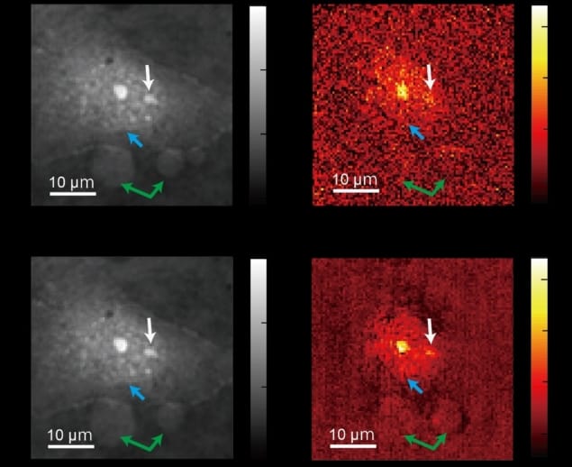
Uncovering the mysteries of how cells function requires advanced imaging techniques to look inside the cells themselves. Research from the University of Tokyo introduces a new microscopy technique that allows us to see cellular structures in much more detail than before, without the need to use any dyes or high-power lasers that may alter the cell.
Taking images inside living cells using only light relies on the very small differences in the phase of the light as it passes through different parts of the cell’s architecture. Creating more detailed pictures requires the ability to detect even the most subtle of these changes with the highest possible sensitivity. This latest work, published in Light: Science & Applications, unveils ADRIFT-QPI – adaptive dynamic range shift quantitative phase imaging – a technique that promises to push the boundaries of sensitivity further than was previously thought possible.
Taking a second look
ADRIFT-QPI uses two steps to capture both the major features and small details of the cell in one image. The first step employs a phase imaging method that’s already widely used, in which a sheet of light is sent towards the cell and the resulting phase shift of the light provides an initial outline of what the cell looks like.
The key development lies in the second step. Another, brighter, sheet of light is used that mirrors the features of the cell captured in the first step. If the first exposure captured a perfect image, the changes caused by the second light sheet passing through the cell would create a completely blank image. By using a much brighter light source and filtering out all of the major features already detected, this second image captures the fine details that previously would have been drowned out in the first step. Combining these two steps ensures that a much wider range of cellular features can be captured in a single image.
“This is the interesting thing: we kind of erase the sample’s image. We want to see almost nothing. We cancel out the large structures so that we can see the smaller ones in great detail,” explains Takuro Ideguchi, whose research group led this work.
Illuminating the smallest details
The significant increase in sensitivity over conventional phase imaging that this method provides (almost seven times greater) allows even the smallest features, such as viruses, to be seen using just light. “Our ADRIFT-QPI method needs no special laser, no special microscope or image sensors,” says Ideguchi. “We can use live cells, we don’t need any stains or fluorescence, and there is very little chance of phototoxicity [an effect often seen when high-powered laser light damages or kills the cells].”
Ultimately, this method could open the door to a closer understanding of the behaviour of small particles on the nanoscale – objects that are hundreds of times smaller than the width of a human hair. Ideguchi offers the following example: “small signals from nanoscale particles like viruses or particles moving around inside and outside a cell could be detected, which allows for simultaneous observation of their behaviour and the cell’s state.”


