Available to watch now, Agilent Vacuum Products, in partnership with IUVSTA, explores the theoretical background of Auger electron spectroscopy and the necessary instrumentation
Want to learn more on this subject?
Auger electron spectroscopy is a well-established surface-sensitive technique. It has been mostly used for the identification of species on sample surfaces, especially to monitor the process of sample cleaning in high vacuum.
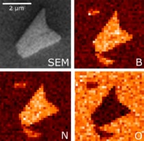
Compared to X-ray photoelectron spectroscopy (XPS), it is much more challenging to gather information on the chemical state of the elements on the surface from Auger spectra. Hence, XPS is a preferred tool for elemental and compositional analysis in many areas of surface science. However, with the advent of nanotechnology, the interest in spatially-resolved elemental information has been raised. In this regard, Auger electron spectroscopy can be easily turned to microscopy, offering very high spatial resolution while keeping the sensitivity to the very surface of the sample. This makes it a unique tool for nanoscale science.
During this webinar, Miroslav Kolíbal will discuss the theoretical background of Auger electron spectroscopy and microscopy, and the necessary instrumentation. In the second part, he will focus on selected applications of Auger spectroscopy utilizing a finely focused electron beam for site-specific Auger analysis.
Want to learn more on this subject?
 Dr Miroslav Kolíbal joined the group of Dr Heike Riel at IBM Zurich Research Laboratory, Switzerland, in 2012, developing high-k dielectric templates for directed III-V nanowire growth. Alongside this he joined a newly established centre of excellence, CEITEC in Brno, Czech Republic, and participated in designing its laboratories. In 2017, Miroslav defended a habilitation thesis at the Faculty of Mechanical Engineering at BUT and became an associate professor in applied physics. He has continued his research on real-time electron microscopy and electron-beam-induced effects at CEITEC in close collaboration with R&D research labs of ThermoFisher Scientific. In January 2020, the successful collaboration resulted in the donation of a dual-beam microscope to his lab.