Researchers in Germany have succeeded in laser printing a special type of stem cell – so-called human-induced pluripotent stem cells (hiPSCs) – for the first time. These hiPSCs are prized for their ability to be differentiated into any type of human cell, a property called pluripotency, plus they can be generated from a patient’s own cells to avoid the risk of an immune response when they are implanted back into the body.
As a result, many scientists believe that printed hiPSCs offer the best option yet for making replacement organs or organ-on-chip systems for personalized drug testing. The problem is that these cells are notoriously fragile and difficult to handle, especially when dissociated into single cells.
“Dissociation is required for printing these cells in high resolution patterns, but it induces programmed cell death (although this can be delayed by adding specific culture medium supplements),” explains Lothar Koch of Laser Zentrum Hannover, lead author of the study published in Biofabrication. “They are thus difficult to print with established techniques such as extrusion, ink-jet printing, acoustic droplet ejection techniques, laser-guided direct write, and laser bioprinting.”
What’s more, continues Koch, the cells’ pluripotency and directed differentiation are affected by environmental factors, such as the composition of the culture media in which they are grown and the bioinks used to print them. They are also sensitive to mechanical forces, such as the shear stresses that can occur during bioprinting processes.
The new laser printing technique developed by Koch and colleagues exploits laser pulses to expel tiny droplets of a bioink containing suspended hiPSCs from a thin layer of bioink deposited on a glass slide. “The main difference with previous approaches, such as extrusion or ink-jet printing, is the absence of a nozzle,” says Koch. “Although this makes preparation and application of the bioink more complex, it does mean that we avoid the high shear forces that usually occur in small nozzles.”
Any 2D pattern and 3D layer-by-layer patterns
The technique combines small droplet printing, down to a few picolitre volumes, with high-viscosity bioink printing and high cell densities of up to 108 cells per millilitre. “Each of these points can be achieved with other printing techniques too, but not in combination,” adds Koch.
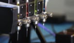
The laser bioprinting set-up comprises the laser and two glass slides. “We coat the top slide with a thin layer of laser-absorbing materials,” says Koch. “This might be a biocompatible metal such as gold or titanium, or a polymer such as triazene or polyamide, or even a hydrogel like gelatine. Then, we deposit the biomaterial to be printed – usually a cell-containing sol – as a second layer on top of the absorption layer.”
The coated glass slide is mounted upside-down above a second slide, and 10 ns laser pulses are then focused through the upper slide into the absorption layer. “This causes a vapour bubble to expand and propel a small volume of the biomaterial towards the lower glass slide,” explains Koch. “By moving the laser and the glass slides, we can print any 2D pattern and also generate 3D layer-by-layer patterns. It is also possible to put substrates or scaffolds on the lower glass slide and print the biomaterial directly onto the substrate or scaffold.”
Survival rate of nearly 100%
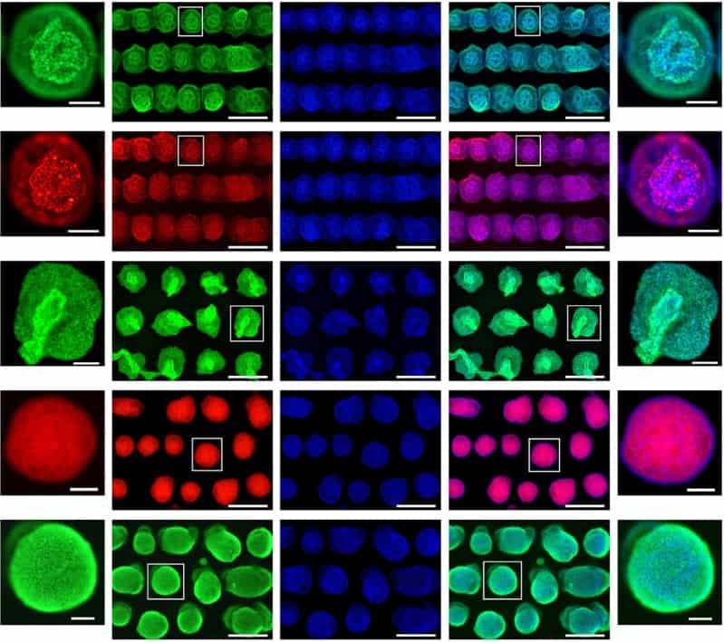
Koch told Physics World that all the cell types tested in these experiments survived this printing procedure, with a survival rate of nearly 100%. The technique also retains the pluripotency of the cells, and allows them to be printed into highly controlled patterns to generate functional tissue substitutes.
The researchers tested a variety of hydrogels as bioinks and culture substrates for printing, and found that fibrinogen, blood plasma, Matrigel and hyaluronic acid were particularly suitable. “Hyaluronic acid is naturally found in the human body; it is generated in the early stage of embryogenesis and is abundantly found in stem cell niche environments,” explains Koch. “It enhances the proliferation of stem cells and supports pluripotency too. It is also good for the laser-printing process and allows us to fine tune the bioink’s viscosity.”
The past, present and future of 3D bioprinting
While Koch believes that hiPSCs are the most promising types of cells for printing tissue or organs, he highlights the need to investigate the process in more detail. “An important question we must ask ourselves is: which stage of differentiation is the optimal one for printing? To answer this, we need to investigate the printability of hiPSCs in all possible stages of differentiation.”
The team, says that it will now be developing a more advanced bioink to print more complex 3D structures. “Another interesting application for printed hiPSCs is to use them to generate cell constructs that mimic embryoid bodies for modelling human development or investigating diseases,” says Koch. “We will be looking to print such embryonal organoid models with our technique.”
- Read our special collection “Frontiers in biofabrication” to learn more about the latest advances in tissue engineering. This article is one of a series of reports highlighting high-impact research published in the IOP Publishing journal Biofabrication.


