A new liver-on-a-chip made from natural collagen could offer a more realistic model for drug screening, as well as for the study of pathological diseases such as liver cancer and cirrhosis. The new device, which is made from self-assembled endothelial cells and two types of collagen in separate layers, remains bioactive for at least seven days.
One of the main reasons that new pharmaceuticals fail clinical trials is the damage they cause to the liver, which metabolizes all drugs ingested into the body. Researchers thus need to develop good in vitro models for evaluating the toxic effects that drugs can have on the liver, known as hepatoxicity. Conventional drug-screening techniques, such as animal testing, are time-consuming and costly, and can also be inaccurate.
A liver-on-a-chip offers an attractive alternative, thanks to its small size, precise architecture, and a controllable biomimetic physiological environment that allows scientists to carry out accelerated bioreactions. A research team led by Wei Sun of Tsinghua University in Shenzhen, China, has now fabricated a new device that more closely mimics the natural physiological environment of the liver, which should enable a more accurate assessment of hepatoxicity (Biofabrication 10 025010).
Close to nature
The new device is designed to replicate the function of liver sinusoids, low-pressure vascular channels that mix oxygenated blood from the hepatic artery with nutrient-rich blood from the portal vein. These channels are flanked by plates of liver cells called hepatocytes, and the region between the endothelial lining of the sinusoid and the hepatocytes is called the “space of Disse”. Plasma from sinusoidal blood can flow almost unimpeded into this space, and the plasma that collects here flows back towards the portal tracts and then into the body’s lymphatic system.
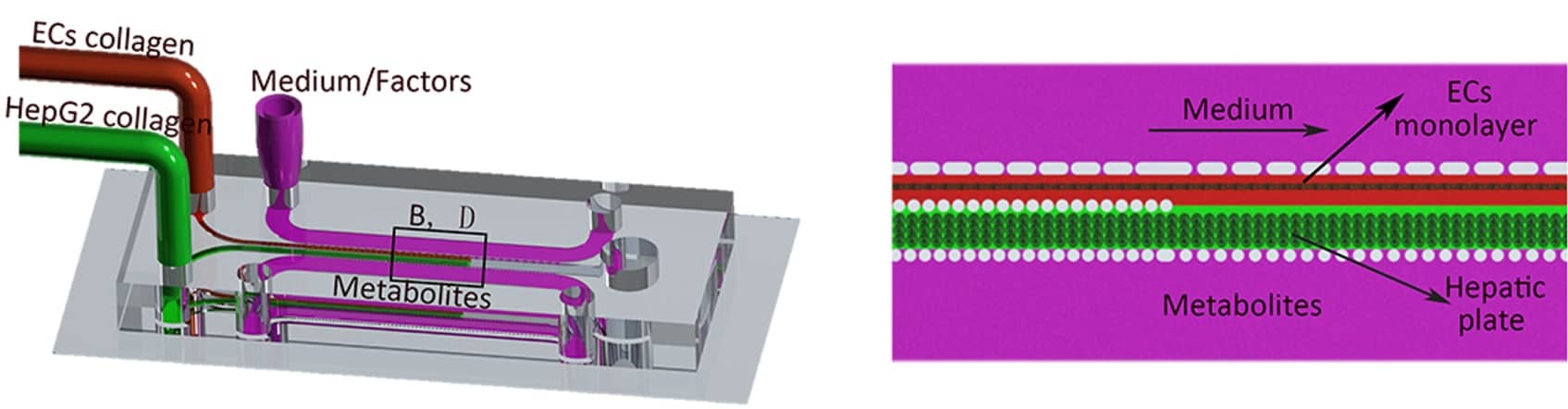
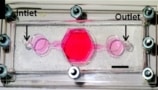
To make their liver-sinusoid model, Sun and colleagues first used standard soft lithography to fabricate a dimethylsiloxane (PDMS) chip with three chambers. Two kinds of natural collagen – one laden with hepatocytes and the other laden with endothelial cells (ECs) – were then simultaneously injected into two of the three chambers. By carefully controlling the flow rate, the researchers were able to form layers of cell-laden collagen with clear boundaries between the two types.
Stimulating self-assembly
“We then injected growth factors into the chamber next to the EC-laden collagen to stimulate the self-assembly of these endothelial cells,” explains team member Shengli Mi. “After roughly two days, the ECs in the collagen formed a monolayer – and this was our liver sinusoid on a chip.”
Since the team’s model is made of natural collagen, it more closely mimics the biological environment in the human body – in which the collagen degrades slowly over time and is gradually replaced by the collagen produced from cells in the liver sinusoid. “This means that the reactions that occur in our device more closely resemble real biological reactions,” says Mi.
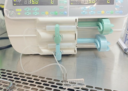
And that’s not all: the team also employed a passive micro-infusion pump rather than a traditional electric one to refresh the medium and to enable continuous nutrition exchange. According to Mi, this yields a cheaper device that’s also capable of high throughputs.
The researchers carried out basic quantitative measurements on the chip, such as cell viability, albumin secretion and urea synthesis, to test out its biomimetic function. “By comparing these functions with those in the presence of different concentrations of a hepatoxic drug like acetaminophen, for example, we could calculate how this drug affects the liver,” Mi told Physics World. “And by setting an allowable cell viability range or functional viability range, we could determine at which dose the drug was safe.”
Studying liver disease
Since cells migrate in the model, it can also be used to analyse the movement of cancer cells to or from the liver – which could help the study of liver diseases such as liver cirrhosis and cancer. The 3D self–assembly described in this study, which is published in the journal Biofabrication, might also come in useful for constructing different kinds of organs-on-a-chip.
Liver-on-a-chip assesses drug toxicity
The team says that it will now be focusing on designing improved passive pumps for its chip, which would allow longer periods of steady nutrition exchange. “What’s more, we will now also be designing a model with a greater variety of liver cells, such as hepatic satellite cells and Kupffer cells, and a more complex system-on-a-chip with biomimetic laminar structure and function for extended applications,” says Mi.
- Read our special collection “Frontiers in biofabrication” to learn more about the latest advances in tissue engineering. This article is one of a series of reports highlighting high-impact research published in the IOP Publishing journal Biofabrication.


