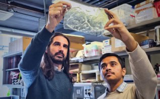
Embedding electronic circuitry inside human tissue has long been a mainstay of science fiction. Now, scientists in the US have devised a way to grow a culture of live tissue over a matrix containing tiny electronic sensors. As well as leading to better tissue cultures for drug testing, the work could also contribute to the development of synthetic replacement organs.
The growth of living tissue with embedded electronic sensors could have a range of biological and medical applications. However, the only option up to now had been to culture the tissue and then to insert electrodes into it. This is undesirable for two reasons. First, a series of electrodes pushed in like needles do not access the tissue in a precise and sensitive manner. Second, inserting electrodes into tissue will inevitably cause damage.
Now, Charles Lieber’s team of chemists at Harvard University has teamed up with tissue engineers at the Massachusetts Institute of Technology and Boston Children’s Hospital to develop a better way of integrating tissue and electronics. Instead of using traditional electrode-based detectors – which deliver weaker signals as they are made smaller – Lieber and colleagues opted for silicon field-effect transistors (FETs) as detectors. FET sensors can be extremely small – in this case made from 30 nm-diameter nanowires – and still give accurate readings.
Non-invasive process
The FETs, together with the interconnecting circuitry, were embedded within a special porous, biocompatible 3D matrix. The researchers then cultured the tissue over the top of this matrix, which created a fine network of FET sensors embedded inside the tissue. “The big difference between our method and the older method is that our method is a non-invasive process,” says Jia Liu, a student in Lieber’s lab and one of three lead authors of a paper on the work. “When we record or stimulate the tissue, we don’t need to use electrodes that puncture through the tissue.”
The researchers tested to see whether the presence of these sensors would have any impact on cell viability over several weeks and found that any effect was minimal. They admit, however, that longer-term studies would be necessary before the technology could be used to create medical implants.
To demonstrate the usefulness of their technology for drug testing, the researchers produced a tissue of cardiac cells integrated with FET sensors. They used the sensors to monitor the effect on the cardiac tissue of noradrenaline, a drug that speeds up the heart rate. They measured a twofold increase in the tissue’s contraction frequency following the application of the noradrenaline.
Synthetic muscles
“This is an excellent paper and the very first example of combining flexible electronics with tissue engineering. Nanowire-based flexible electronics technology could be one of the best approaches to such 3D tissue scaffolds that can be electrically probed,” says Zhenqiang Ma, an expert on epidermal electronics at the University of Wisconsin-Madison. Ma suggests that the technology could be particularly valuable for producing synthetic versions of tissues such as muscles and neurons that involve electrical signals in their functions.
In the near future, the researchers believe that the work’s applicability is likely to be confined to improving tissue cultures for drug testing. Nevertheless, Liu agrees that, in the long term, the contribution to the quest to produce synthetic versions of body parts could be significant. He explains that researchers in this field already use extracellular matrices of the type used here to culture synthetic tissue. “In the past, however, this tissue scaffold has been a passive material that just supports the cells as they grow,” says Liu. “But right now, we have made a nanoelectronic tissue scaffold that not only supports the growth of the cells, but can also monitor their functionality.”
The research is published in Nature Materials.


