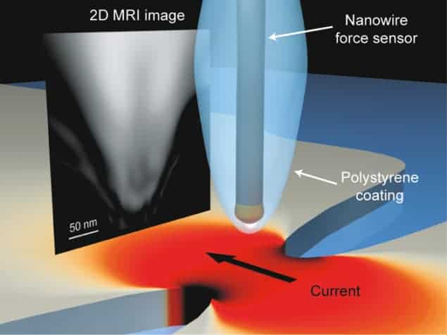
Magnetic resonance imaging (MRI) at spatial resolutions of just 10 nm has been achieved for the first time. Developed by researchers at the University of Illinois at Urbana-Champaign in the US, the technique could be particularly useful for imaging biological samples. If further improved, it could even be used to image viruses and protein macromolecules.
MRI is based on nuclear magnetic resonance (NMR) and is a powerful tool that allows scientists to study the chemical composition of many different materials. This includes living tissue and as a result MRI has become a powerful diagnostic tool in medicine. The technique works by measuring the response of the nuclear magnetic moments – or spins – of a sample to external magnetic fields and electromagnetic radiation. But because the individual nuclear moments are tiny, MRI signals from small samples are weak and can be easily swamped by noise. As a result it has proven to be difficult to do MRI at spatial resolutions of less than about 1 mm, except under special circumstances.
This latest work was done by Raffi Budakian and colleagues, who attached the sample to be analysed – a tiny piece of polystyrene – to the tip of a silicon nanowire mechanical resonator. This is a small plank of silicon roughly 15 µm long and 50 nm wide. They then place this nanowire over a metal constriction 240 nm wide and 100 nm thick. By passing high-frequency electric currents through the constriction, they are able to generate the intense magnetic fields needed to do MRI.
Tiny vibrations
The team then oscillates this electric current through the constriction to generate a magnetic-field gradient that alternates at the same frequency as the nanowire vibrates. The interaction between the magnetic moments in the sample and the alternating inhomogeneous magnetic field produces tiny vibrations of the nanowire that can then be measured using an optical interferometer included in the set-up.
The Illinois team was able to successfully image hydrogen nuclear spins in the polystyrene sample using its method and obtained a 2D projection of the hydrogen density in the material with a spatial resolution as small as 10 nm.
“Another important result of our research is that we have demonstrated a new magnetic resonance protocol that allows us to apply NMR techniques to encode so-called spin noise,” explains Budakian. “That is, we encode information in the statistical fluctuations of all the nuclear spins in a sample rather than in their thermal spin polarization – as is usually the case.”
“Our technique in fact uses well established methods in MRI,” Budakian says. “Fourier-transform imaging is routinely used in MRI and is a very efficient sample-imaging technique, but the main difference in our new method is that we encode information in the spin noise rather than in the thermal polarization.”
According to the researchers, the technique could come in handy for imaging biological samples. “Our near-term goal is to achieve even higher spatial resolution and begin imaging virus particles,” adds Budakian. “We would ideally like to tomographically image virus particles in 3D and, with sufficient improvement, might even be able to image macromolecules such as proteins in the future.”
The work is reported in Physical Review X 10.1103/PhysRevX.3.031016.
- This article first appeared on nanotechweb.org


