
Biomaterial investigators know that the mechanical stretching of a scaffold containing cells modifies the cells’ behaviour. This effect is being studied and used by research groups worldwide to optimize the fabrication of collagen scaffolds – biomaterial constructs based on collagen that can carry cells and be implanted in the body.
Diego Mantovani and his group at Laval University (LBB) are well aware of this issue and are aiming some of their research in this direction. In a recently published article, they explore the idea of identifying the correct “work out” or stretching that cells in 3D collagen scaffolds must follow to optimize their effect on the collagen. Their results show that an incremental frequency of strain can improve the properties of the scaffold in which the cells are growing (ACS Biomater. Sci. Eng. doi: 10.1021/acsbiomaterials.7b00395).
Making the cells fabricate the scaffold
At the recent Advanced Materials for Biomedical Applicationsconference in Ghent, Mantovani gave a fascinating talk about the collagen tubular constructs that his team is producing for vascular tissue engineering. However, the fibrillary arrangement of collagen scaffolds is not being carried out by the researchers directly, but by the cells themselves.
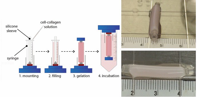
Cells in a collagen–gel solution are doing the work of remodelling the extracellular matrix protein produced by themselves, together with the exogenous collagen provided by researchers. All this happens during one to two weeks maturation inside a custom-designed bioreactor, a closed system that allows the growth of cells in the desired conditions.
These systems allow the induction of a symphony of stimuli – including stretching of the material that the cells grow on, or the flow of liquid – which are detected by the cellular sensors and modify their metabolism. One of the effects that these stimuli have on the cells is to motivate them to alter the material architecture and to improve mechanical properties, as shown in an earlier publication (Ann. Biomed. Eng. 45 1496).
Are in vitro and in vivo mechanical stimuli similar?
There are still many conditions to optimize but, if we focus purely on the mechanical stimuli, we can ask: Is a constant mechanical stimulation representative of or relevant to what actually happens in the native blood vessels? Are we making the cells “work out” correctly?
A strain with incremental frequency is indeed more representative of what occurs in the blood vessels in vivo than a constant strain. In addition, it might provide new insights as to how mechanical strain affects cell behaviour in a 3D environment in vitro. To compare and understand the behaviour of the cells under the different conditions, the researchers monitored their shape, orientation and expression, and studied the mechanical properties of the scaffolds.
A gradual increase improves remodelling
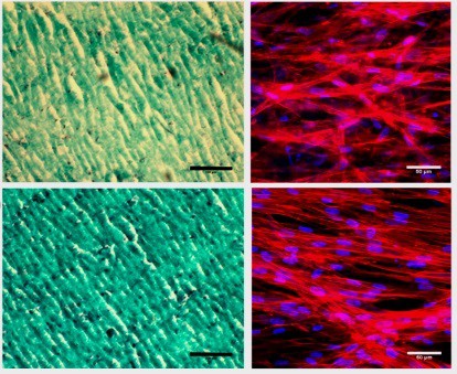
As expected, cells showed different behaviour under the different conditions. The gradual increasing strain promoted a higher alignment of cells and their nuclei when compared with the other conditions. Moreover, it even improved the remodelling of collagen, showing a more compact and aligned structure. Importantly, this alignment occurred in the direction of the strain, as seen in native blood vessels.
However, even though the cells improved the remodelling of the tissue under strain, the expression of proteins related to tissue remodelling was higher in the static control. This fact, together with similar observations reported in other articles, could be accounted for by the “desensitization of the cells over time to cyclic mechanical stimulus” since “no remodelling is seen in the human vasculature unless changes in mechanical cues or injuries are sensed”, the authors suggest. Furthermore, the cells under gradual strain within the scaffold exhibited improved mechanical properties, providing closer characteristics to those observed in the native blood vessels.
The study demonstrates that the use of an incremental frequency in the strain strengthens the resemblance between native blood vessels and in vitro developed scaffolds, reducing the gap between them.
Although this provides some answers to current models in vitro, it nonetheless poses more questions: Would we expect the same outcome by incrementing the intensity of the strain? Would there be a synergic effect between the intensity and the frequency? Could we observe similar behaviour when combining with the stimulus of the flow?
While these questions remain unanswered, Mantovani and his group at LBB will be making cells “work out” to provide future answers.


