
Scientists in the US have conducted the first clinical tests of a new diagnostic device called MasSpec Pen. The “pen” is able to detect differences between healthy and cancerous tissue in a matter of seconds, to help surgeons decide which sections must be removed during pancreatic cancer surgery. The researchers, from The University of Texas at Austin and Baylor College of Medicine, describe their research in Proceedings of the National Academy of Sciences.
Cancer of the pancreatic duct is one of the most lethal types of cancer, with less than one in 10 patients surviving five years after diagnosis. To ensure that patients have the best chance of survival, doctors must surgically remove those sections of the pancreas affected by cancer. However, distinguishing healthy cells from cancerous tissue is a difficult task.
The current method for differentiating tissue types involves examining tiny frozen samples removed from the pancreas during surgery. But this frozen section analysis is time-consuming and can be inaccurate.
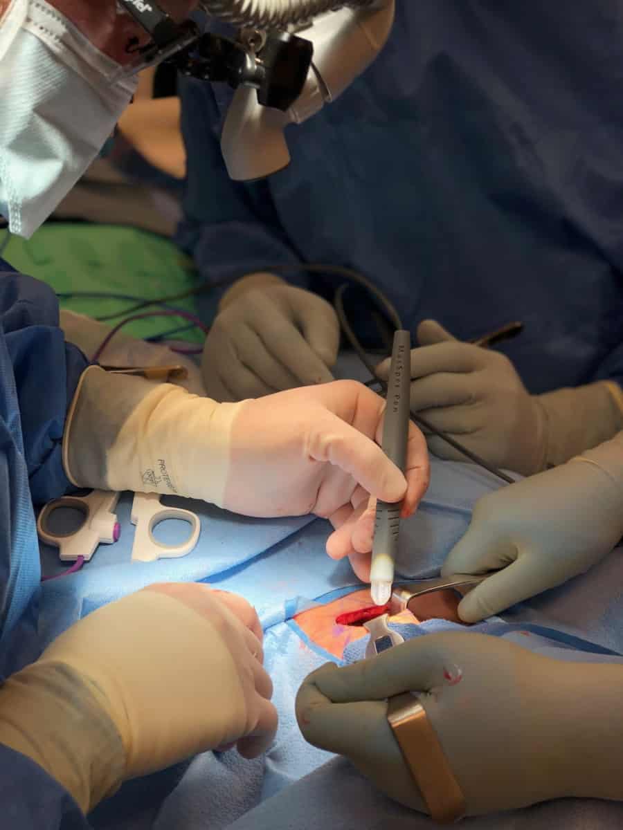
In 2017, the team – led by Livia Eberlin at UT Austin and James Suliburk at Baylor College – developed the MasSpec Pen as a complementary tool to frozen section analysis. This technology provides surgeons with additional molecular information in vivo to guide decision-making, explains Mary King, first author of the present study.
Surgeons can use the pen to extract biomolecules from the surface of pancreatic tissue. These molecules then feed into a connected mass spectrometer, which detects differences between cancerous and healthy cells from their molecular profiles. Tissue classification using the MasSpec Pen takes about 15 s – one hundred times faster than using frozen section analysis.
In their recent study, the researchers first used the MasSpec Pen in the laboratory to analyse 157 banked samples of human pancreatic tissue. Using the molecular profiles of these samples, they trained a statistical model to classify tissue as either cancerous or healthy.
They then transferred the setup to the operating room at Baylor St. Luke’s Medical Center in Houston. Here, the researchers analysed tissues in vivo and immediately after removal during 18 pancreatic cancer surgeries. They found that predictions from their statistical model were accurate in more than 93% of the samples analysed. They also observed that training their classification algorithm with both banked samples and samples collected from the operating room boosted the agreement between their predictions and post-surgery tissue analysis.
“These results show the technology works in the clinic for surgical guidance,” says Eberlin. “Surgeons can easily integrate the MasSpec Pen into their workflow, and the initial data really support the diagnostic accuracy we were expecting to achieve.”
The scientists are currently expanding their study with more patients and are also testing the MasSpec pen in breast cancer and ovarian cancer surgeries. King also emphasizes that they are working to improve the useability of their device in hospitals. “One of our future goals is adapting the MasSpec Pen setup with smaller and more mobile mass spectrometers to make it even more hospital and operating room-friendly,” she tells Physics World.


