This year, the Physics World team selected a medical innovation as the Breakthrough of the Year: the development of a digital bridge that restores communication between the brain and spinal cord, enabling a man with paralysis to stand and walk naturally. We also reported on several other neural engineering advances, including a neuroprosthesis that restores communication to those who cannot speak and an award-winning implant that could help regulate blood pressure in people with spinal-cord injuries.
And that’s just one example of the impact of physics-related research on the healthcare sector. In 2023, we wrote about a host of medical physics and biotechnology advances, from photon-counting detectors that produce high-quality images with less contrast media to hydrogels that help grow new brain tissue to shoot-through FLASH proton therapy. Here are a few more highlights that caught our eye.
Novel takes on nuclear medicine
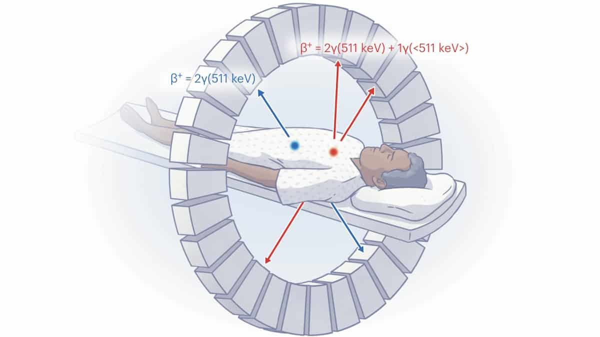
Among the many developments in positron emission tomography (PET) technology announced this year, a research team headed up at Memorial Sloan Kettering Cancer Center and Complutense University of Madrid devised a novel image reconstruction method that enables in vivo imaging of two different PET tracers simultaneously. This “multiplexed PET” technique, which increases the amount of information attainable during a single scan, can be implemented on preclinical or clinical PET systems without having to modify the hardware or image acquisition software.

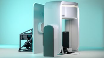
Aiming to meet the ever-increasing clinical demand for PET scans, researchers at Ghent University in Belgium are developing a walk-through total-body PET scanner. Their proposed upright imaging system, which looks a bit like an airport security scanner, is expected to be both cheaper and quicker to use than standard PET instruments.
And researchers at UC Davis used total-body PET to perform first-in-human immunoPET imaging of T cell biodistribution in three healthy individuals and five patients recovering from COVID-19. Quantification of immune cell distribution and kinetics in humans can shed light on how the immune system responds to viral infections, and help researchers develop new vaccines and improved treatments.
Radiotherapy for the future
The introduction of MR-guided radiotherapy, which uses MRI to visualize tumours and surrounding organs with high accuracy while the patient is on the treatment table, enables clinical advances such as real-time plan modification and imaging of moving tumours during treatment delivery. But integrated MR-linac systems hold potential to do a lot more.
Researchers at the University Hospital of Zurich investigated an approach called adaptive fractionation, which exploits inter-fraction motion (rather than simply compensating for it) by adjusting the prescribed dose according to the distance between the tumour and organs-at-risk (OAR) on each day. In other words, a patient is prescribed a higher radiation dose on days when their tumour–OAR separation is large and a lower dose on days when this separation is small.
Elsewhere, a team at the University of Toronto’s Sunnybrook Health Sciences Centre studied the use of diffusion-weighted imaging (DWI) on an MR-linac to improve treatment of the aggressive brain cancer glioblastoma. The idea here is to use DWI to identify regions of treatment-resistant tumour and deliver higher doses to these targets.

Another technology to keep a close eye on is the introduction of upright radiotherapy, pioneered by Leo Cancer Care. Back in January we reported on a patient positioning system that allows cancer patients to receive radiotherapy whilst sitting upright – in contrast to having to lie on their back – a position that should reduce organ movement during treatment and may also be more comfortable for the patient. Since then, several studies have confirmed the benefits of this upright approach for various tumour types, orders have been placed, and the positioning system is now pending 510(k) regulatory clearance for clinical use in the USA.
Wearable wonders
Each year we see the emergence of ingenious wearable devices for countless new healthcare monitoring and diagnostic applications; and 2023 was no exception.

For starters, a team headed up at The University of Texas at Austin created an ultrathin, stretchable electronic tattoo that provides continuous cardiac monitoring. Attached to the chest via a medical dressing, the e-tattoo could detect early signs of heart disease outside of the clinic. Meanwhile, a wearable ultrasound transducer developed at the University of California San Diego could be used to monitor patients with serious cardiovascular conditions, as well as to help athletes keep track of their training.

Medical physics and biotechnology: our favourite research in 2022
Other novel wearables reported this year include a ring device that accurately gauges how intensely its wearer is scratching their skin, a miniaturized ultrasound scanner that may provide earlier detection of breast cancer, and earbud biosensors that continuously and simultaneously measure the electrical activity of the brain and levels of lactate in sweat.
Finally, researchers from the Medical University of Vienna designed a prototype MRI coil that can be worn like a sports bra. The so-called BraCoil is a vest-like receive-only coil array made of flexible coil elements that enables 3 T MR imaging of patients in both supine (lying on their back) and prone (lying on their front) positions. Designed to improve comfort, and reduce preparation and acquisition time, the BraCoil also produced an up to three-fold improvement in signal-to-noise ratio compared with standard coils.
Physics World‘s coverage of the Breakthrough of the Year is supported by Reports on Progress in Physics, which offers unparalleled visibility for your ground-breaking research.


