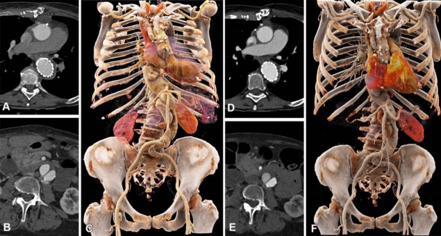
Photon-counting detector CT (PCD CT) is heralded as a major technological advance in CT imaging. Photon-counting systems measure each individual X-ray photon, providing increased image resolution and a reduction in radiation dose. Researchers from the University of Zurich have now demonstrated that the technique can reduce the amount of iodinated contrast media required for CT angiography (CTA) by 25%, while producing comparable image quality to conventional CTA.
CTA is the standard imaging exam used to assess cardiovascular disease and for follow-up after surgical interventions. The ability to use less contrast reduces the risk of contrast-induced acute kidney injury in patients with kidney disease, diabetes or arterial hypertension. As well as making CTA exams safer for patients, reducing the use of iodinated contrast also benefits the environment, as iodine breakdown products have harmful environmental effects.
The introduction of PCD CT has energized CT research, with numerous studies examining how the technology may change the use of CT and improve its diagnostic capability. In September 2021, the Siemens NAEOTOM Alpha became the first commercial photon-counting CT system to achieve 510(k) clearance from the US Food and Drug Administration (FDA). The FDA stated that this represented “the first major new technology for computed tomography imaging in nearly a decade”.
Making CTA safer
In their prospective study, described in Radiology: Cardiothoracic Imaging, the University of Zurich researchers aimed to develop and evaluate a low-volume contrast media protocol for thoraco-abdominal CTA using PCD CT. The study included 100 patients who underwent CTA with PCD CT of the aorta in the chest and abdomen, and had previously undergone a CTA exam with conventional energy-integrating detector (EID) CT at equal radiation dose.
The EID CTA exams used 70 ml of iodinated contrast media: delivered as a 40 ml bolus of contrast, followed by 60 ml of a 1:1 mixture of contrast and saline, and then a saline flush. This protocol was also used for PCD CT angiography scans of 40 patients (group 1). The researchers then imaged 60 patients (group 2) with PCD CT using a low-volume contrast media protocol: a bolus of 30 ml contrast, followed by 45 ml of 1:1 mixture of contrast and saline, and a saline flush. This equates to a total contrast volume of 52.5 ml – a 25% reduction compared with the first group.
Principal investigator Kai Higashigaito and colleagues report that for group 1 patients, PCD CT improved the overall image quality. “PCD CTA of the aorta demonstrated higher objective and subjective image quality compared with third-generation EID CT at equal contrast media volume and matched radiation dose,” they write.
To determine the optimal energy of virtual monoenergetic imaging (VMI) for PCD CT-based CTA, the researchers assessed the images from group 1 for objective qualities such as contrast-to-noise ratio (CNR) and subjective qualities such as image noise. They observed the highest objective image quality at 40 keV and the highest subjective image quality at 60 keV. Thus they selected 50 keV as the ideal energy of VMI for CTA of the aorta in the group 2. In these patients, who received 25% less contrast, PCT CT produced noninferior images to EID CT.
PCD pioneers
Co-author Hatem Alkadhi tells Physics World that University Hospital Zurich was the first clinical site in the world to install a PCD CT system for clinical use, in April 2021. The scanner is now routinely used for all cardiovascular CT examinations, including CTA, of all body regions.
“We are currently conducting various research studies with PCD CT, mainly in the field of cardiovascular imaging,” he explains. “As an example, we are currently developing a low-volume contrast media protocol for coronary CT angiography. Our initial results indicate that we can reduce the amount of administered contrast media by 40% as compared to our current standard, while still obtaining diagnostic image quality.”

Photon-counting CT promises a new era of medical imaging
Alkadhi says that the team is also studying the ultrahigh-resolution mode of PCD CT, which enables image reconstruction with very high spatial resolution. This technique improves coronary artery and coronary plaque imaging, he says, but also improves coronary stent imaging, with a better visualization of the in-stent lumen.
In addition, the researchers are investigating the spectral capabilities of their dual-source NAEOTOM Alpha scanner, which enables dual-energy-based calcium–iodine separation in the arteries. They are using an algorithm called PureLumen to subtract contributions from calcified plaques from the vessel wall image. “Thus, a major shortcoming of coronary CTA – the overestimation of coronary stenoses because of blooming artefacts from dense vessel wall calcifications – can be overcome with this very exciting technique,” Alkadhi explains.


