Welcome to the latest in a new series of sponsored articles showcasing some of the latest white papers and webinars from physics-based businesses around the world
This time we are featuring webinars from Park Systems and WITec, as well as a white paper from Lake Shore Cryotronics.
Advances in microscopy
![]()
Piezoelectric force microscopy (PFM) is a form of scanning probe microscopy (SPM) that lets researchers image and manipulate piezoelectric and ferroelectric domains in materials. In this webinar, members of the technical-marketing team at Park Systems introduce the principles behind the technique, which involves engaging a sample surface with a sharp conductive SPM probe and then applying an AC current to the probe’s tip to deform the surface via a piezoelectric force. The PinPoint variant of PFM, specific to Park Systems’ atomic-force microscopes, lets you image samples with higher spatial resolution and with no lateral force. The webinar, PinPoint Piezoelectric Force Microscopy, also reviews the technique’s potential for investigating the electric domain structures (such as polarity) in piezoelectric and ferroelectric materials.
Atomic force microscopy (AFM) is a powerful tool in nanotechnology that has been widely used to study collagen, which is the most common protein in mammals and is found everywhere in connective tissues, including bone, skin, and muscle. Conventional techniques to characterize these fibrils are mainly based on AFM force-volume spectroscopy, which collects force-distance curves at each pixel to calculate material properties. However, these techniques are slow and it can take hours to acquire an elasticity map, which is one reason why Park Systems has developed the PinPoint Nanomechanical Mode. At least 100 times faster than traditional techniques, it can create a map in minutes, with a correlated topography image that reveals the position and orientation of the sample. In this webinar, In-Air and In-Liquid AFM Imaging with PinPoint Nanomechanical Mode, staff at Park Systems discuss this new mode, which can create quantitative maps of the mechanical properties of various materials, ranging from hard disks to soft tissues, including collagen fibrils.
Scanning ion conductance microscopy (SICM) is a variant of scanning probe microscopy (SPM) that allows researchers to determine the surface topography of samples at nanometre range in a non-destructive manner and under in situ conditions. Scanning electrochemical microscopy (SECM), meanwhile, is another SPM technique, this time letting you study the local electrochemical phenomena at various materials interfaces in liquids. SICM, which uses the increase of access resistance in a nanopipette placed in an electrolyte solution, monitors the ionic current flowing in and out of this probe – a flow that is hindered as the tip closes in on a sample surface. In SECM, in contrast, an electrode tip is used to acquire spatially resolved electrochemical signals over a region of interest, with the 2D raster-scan information yielding images of surface reactivity and information on the rates of chemical processes. In this webinar, Scanning Ion Conductance Microscopy (SICM) and Scanning Electrochemical Microscopy (SECM), applications staff at Park Systems explain the basics of both the techniques and discuss their applications in analytical chemistry and electrochemistry.
Material matters

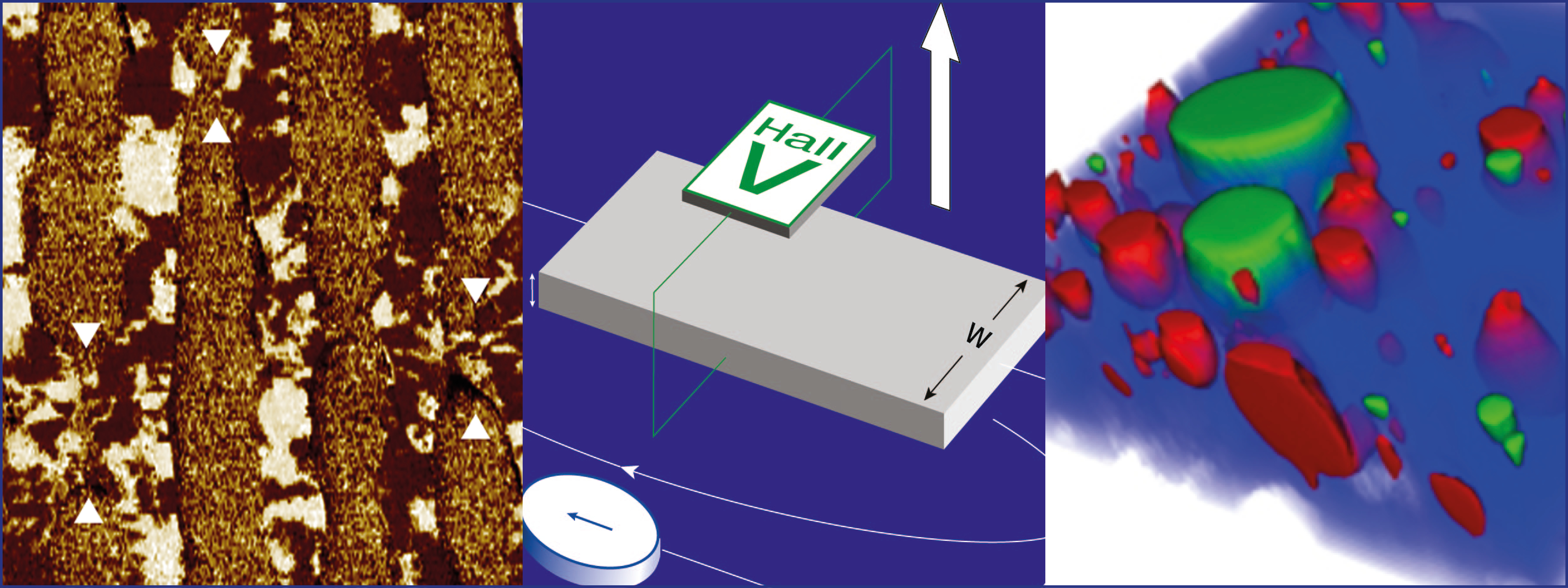
The Hall effect is not just a key phenomenon in condensed-matter physics – it’s also a vital way for characterising the material properties of semiconductors. In the 88-page Hall Effect Measurement Handbook white paper, Lake Shore senior scientist Jeffrey Lindemuth provides both new and experienced material researchers with a comprehensive guide to the theory of Hall measurements. It includes methods to measure the resistivity and the Hall coefficient of materials, major sources of measurement errors, ways to minimize the effects of these errors, and more besides.
Seeing molecules in 3D
Confocal Raman imaging is a powerful, versatile and ever-more common microscopy technique, which can quickly identify the molecules in a sample and visualize their distribution in 3D space. To find out more about this non-destructive and label-free chemical imaging method, which has big potential in many different fields, why not view the webinar The Analytical Power of Correlative Raman Imaging: New Developments, Tools and Applications by Miriam Böhmler and Ute Schmidt. As senior application scientists at WITec, they will introduce the basic principles of Raman microscopy, explain the associated hardware and software, and describe several of its variations – including relevant application examples. They then introduce correlative microscopy and provide details of Raman-AFM and Raman-SEM (RISE) microscopy too.