You might expect that an opaque material will always hinder the transmission of light by absorbing or scattering any light that is shone on it. But by carefully preparing a beam of laser light, physicists in the Netherlands have managed to use scattering in opaque materials to focus the light to an intensified point. Their technique could be used to obtain optical images of biological samples that are hidden beneath layers of opaque tissue (Opt. Lett. 32 2309).

Optical microscopy and spectroscopy both rely on the controlled transmission of light through a sample. But if a sample is covered by an opaque layer – or indeed if a sample itself is opaque – the amount of light randomly scattered can render these techniques next to useless.
Allard Mosk and Ivo Vellekoop from the University of Twente, however, claim that they can not only circumvent the problems of an opaque medium but can actually exploit its properties to focus light to a point over a thousand times brighter than it would be when it is scattered normally.
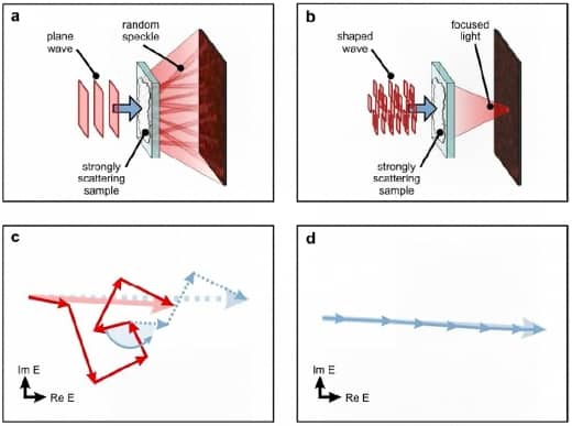
The researchers start by expanding the diameter of the beam from a laser using a lens, and then split the cross-section into a number of segments by passing the beam through the pixels on a liquid crystal display (LCD). After focusing this expanded beam back to its normal diameter, they shine it through an opaque sample onto a digital camera.
The crucial part of Mosk and Vellekoop’s technique is their computer program, which reads the intensity of the light hitting the camera and makes corrections to the LCD’s pattern to make this intensity as large as possible. For example, if one beam segment scatters through the sample in such a way that it interferes destructively with the rest of the beam when it arrives at the camera, the program adjusts that segment’s propagation before it reaches the sample using a phase modulator. When every segment’s phase has been optimized to interfere constructively, the brightest possible image is obtained (See Opaque lens).
Mosk and Vellekoop tested their technique for several opaque samples, some of which turned out to better at focusing than others. A fresh flower petal, they found, focused the beam to an intensity about 60 times greater than the normal scattered beam. On the other hand, titanium dioxide – a white pigment and one of the most strongly scattering materials known – could intensify the beam more than a thousand times over.
In practice, such an intense beam could be scanned over a biological sample to image it in a similar manner to a scanning electron microscope by using any opaque layers of tissue covering it as the “lens”. However, the technique would still require a camera or other detector behind the sample to read the intensity for optimization. “We are starting to work on optimization using local nanoscale probes that can be put inside tissue,” Mosk told physicsworld.com.


