Recent studies have suggested that lead halide perovskites may end the search for the ideal X-ray photoconductor. Common solution-process protocols for fabricating thin films of these materials, however, are difficult to scale to the large areas required for X-ray detectors. In work recently published in Nature Photonics, a team of German researchers has presented an alternative, physical method of processing perovskites to solve this issue. The wafers produced using this sintering technique showed comparable performance to commercially available X-ray photodetectors.
The search for an ideal X-ray photoconductor has led researchers to hybrid organic-inorganic perovskites (HOIP), a class of materials that is already a major target for use in photovoltaics, light-emitting diodes and lasers. Current commercial detection systems based on materials like amorphous selenium and cadmium telluride are limited by low absorption coefficients and stability issues at high energies. HOIP materials, however, have the intrinsic ability to effectively absorb high-energy radiation because their composition includes heavy metal and halide ions. Perovskite X-ray detectors may therefore be a more effective alternative to existing detecting systems.
To date, much of the research has focused on solution-processing of perovskite thin films. While this is an efficient method for producing samples in the sub-micron regime, producing thicker layers, especially over large areas, is extremely difficult. This problem has so far hindered the development of perovskites for medical applications like X-ray detectors.
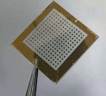
To get around this problem, first author Shreethu Shrestha and colleagues at Friedrich-Alexander-University Erlangen-Nürnberg in Germany, in collaboration with researchers at Siemens and the Bavarian Center for Applied Energy Research (ZAE Bayern), produced layers of perovskite using the physical method of sintering. Squeezing methyl ammonium triiodide perovskite (MAPbI3) powder in a hydraulic press for just five minutes, the researchers formed compact layers, or wafers, that were more than a centimetre in diameter. Depending on the amount of powder used, the resulting wafers ranged in thickness from 200 μm to 1 mm.
According to team member Gebhard Matt, also at Friedrich-Alexander-University Erlangen-Nürnberg, this was remarkable because such a sintering process is not possible for covalent semiconductors like silicon, which lack the plasticity of perovskites. As well as producing large-area detectors, sintering also allows the perovskites to be fabricated at room temperature, which makes the procedure simpler and cheaper than solution-processing.
Scanning electron microscopy (SEM), x-ray diffraction and photoluminescence confirmed that the crystallinity of the material was preserved after the sintering process. The grain boundaries of the microcrystals remain well defined following sintering, but, says Matt, “the surface of the wafers is as smooth as the surface of the cylinder in the hydraulic press”.
The group tested the X-ray performance of their wafers against a commercial cadmium telluride (CdTe) system, Timepix, which is the current state-of-the-art system for X-ray and gamma-ray detection. The group found that their MAPbI3-based detector had sensitivity and conversion values comparable to the Timepix system. CdTe is expensive, and only a few companies worldwide can produce the crystals, so even though the perovskites did not perform better than Timepix, their simpler preparation process makes them an attractive alternative.
Still, the researchers report that much work must be done prior to implementation of perovskite-based X-ray detectors. One major issue they encountered in their device was the presence of a high and unstable dark current. To fix this, the researchers now plan to explore carrier-selective electrodes to improve the performance still further.
More details can be found in Nature Photonics DOI: 10.1038/nphoton.2017.94.