
Propagation-based phase-contrast CT (PB-CT) is an advanced X-ray imaging technology that can generate higher quality diagnostic breast images than absorption-based CT (AB-CT), at a glandular radiation dose comparable to, or lower than, conventional mammography and digital breast tomosynthesis (DBT). The technology is currently limited to use with synchrotron light sources, but the evolution of compact light sources may make clinical application feasible, improving the detection and diagnosis of breast cancer.
A multidisciplinary collaboration – including scientists from the University of Sydney, University of Melbourne, Monash Health, Maroondah Breastscreen and the Australian national science research agency CSIRO – is working on the clinical application of PB-CT using the Imaging and Medical Beamline at the Australian Synchrotron. The researchers have now optimized the technique using 12 mastectomy samples that included different tumour types or benign lesions. They showed that PB-CT achieved significantly higher image quality than AB-CT and demonstrated that substantially lower doses could be used with PB-CT (Acad. Radiol. 10.1016/j.acra.2020.01.009).
Phase-contrast imaging, which exploits both the refraction and the absorption of transmitted X-rays, offers potential to overcome the limitations of current breast imaging modalities. The 3D images produced by DBT reduce the tissue superimposition effects of 2D mammography, but have lower sensitivity in detection of calcifications. Breast MRI has higher sensitivity than mammography, but lower specificity. It is also a highly expensive examination. Breast CT, meanwhile, visualizes mass lesions better than mammography, but underperforms with respect to depiction of microcalcifications and has poorer spatial resolution. In addition, its radiation dose is the highest of the breast imaging modalities.
PB-CT, one technique for phase-contrast imaging, is based on free-space propagation and the use of phase-retrieval algorithms to fully use the refraction information. The PB-CT method measures the phase shift as intensity modulation at the detector by simply positioning the detector a few metres from the object. Unlike other phase-contrast imaging techniques, it does not require any special X-ray optical elements in order to render the X-ray refraction visible. The only key requirements for PB-CT are a long propagation distance between the object and the X-ray detector, and the use of highly spatially coherent incident X-rays, produced by synchrotrons or compact X-ray sources.
The group previously demonstrated that PB-CT could reconstruct images with high quality and high diagnostic value, with a dose comparable to that of 2D mammography. In this latest study, led by Patrick Brennan, the team employed 32 or 34 keV X-ray beams from the synchrotron to scan the mastectomy specimens using PB-CT and AB-CT techniques under varying conditions.
The researchers collected images at two sample-to-detector distances: 0.19 m to represent an AB-CT scan and 6 m for a PB-CT scan. All AB-CT images were collected at a “standard” mean glandular dose of 4 mGy, using 2400 projections with 0.075° angular steps. PB-CT images were collected at both 4 mGy (2400 projections with 0.075° angular steps) and 2 mGy (1200 projections with 0.15° angular steps). The team used an ionization chamber to measure the photon fluence rate and the corresponding rate of the surface absorbed dose to air at the ionization chamber plane.
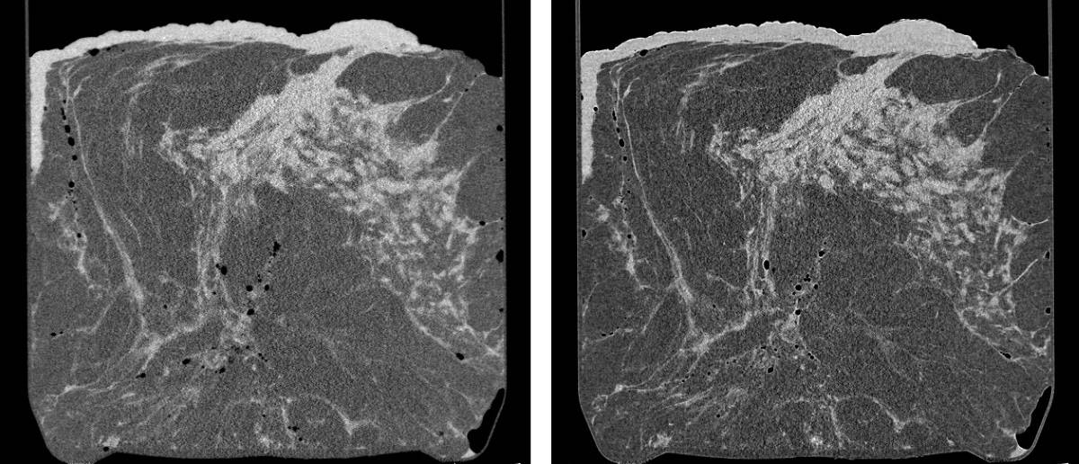
After three radiologists selected the best quality AB-CT image set for each mastectomy specimen, 11 radiologists independently compared the overall image quality in PB-CT images, prepared in axial and sagittal planes, with the corresponding AB-CT images. They evaluated lesion sharpness, visibility of calcifications, image noise, perceptible contrast, visible artefacts and normal tissue interfaces.
The radiologists reported that PB-CT images acquired at both standard and low dose were of significantly higher image quality than the AB-CT images. The researchers also determined that PB-CT images obtained at 32 keV and reconstructed using half phase retrieval (rather than full phase retrieval) had the best overall image quality.
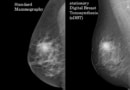
Stationary digital breast tomosynthesis increases diagnostic accuracy
First author Seyedamir Tavakoli Taba tells Physics World that the team will soon start a receiver operating characteristic (ROC) study to compare the diagnostic efficacy of PB-CT with conventional breast imaging techniques, such as mammography and DBT. To date, the researchers have scanned over 75 fresh breast mastectomy samples and anticipate scanning another 50 before launching the first clinical trial, planned for early 2021.
“Our plan is to establish a world-first mammographic PB-CT clinic at the Australian Synchrotron in three to four years, to be used mainly for staging and treatment options,” says Taba. “The widespread clinical implementation of PB-CT can be delivered via commercially available compact X-ray sources in the future. This will allow PB-CT to be widely translated into specialist cancer care facilities across Australia and overseas.”


