Biopsy: an examination of tissue removed from a living body to discover the presence, cause or extent of a disease.
This clinical definition obscures much of a patient’s experience during and after a biopsy. Local tissue injury, bleeding, inflammation, stress and elevated risk of metastasis are not uncommon.
What if there were a less invasive method for performing a biopsy, especially for potential cancer diagnoses? And what if this method could provide information about tumour heterogeneity that conventional biopsies cannot?
Researchers from Tel Aviv University, Herzliya Interdisciplinary and Technion may have an answer. They developed a molecular biopsy technology that can extract RNA and proteins critical to characterizing tissues and applied their approach to tissues extracted from mice (Sci Reports 10.1038/s41598-019-51634-7).
A molecular cartography technology
The researchers’ system uses electroporation – the application of pulsed electric fields to tissues – as a type of molecular biopsy that could be performed even when the precise locations of tumours in a tissue or organ are unknown.
The technique works in two stages. First, researchers apply short electric field pulses at high voltages to make the membranes of cells in tissues vulnerable to external influences. Next, they use longer-duration pulses at low voltages to extract molecular components of interest from these cells. Electroporated molecules are then removed from the tissue using a solvent or other fluid, and pre-existing methods used to analyse molecules of interest.
While the technique sounds simple in principle, a lot of work goes on behind the scenes to optimize and validate the method. Extracting delicate proteins and RNA requires researchers to manage dynamic variables, including electric field strength, pulse duration, pulse number and pulse frequency, to name but a few. In the current work, the researchers performed numerical modelling and related studies to optimize parameters and choose which parameters they would use. Then, they applied their technique to study the molecular signatures of excised mouse livers, kidneys and liver tumours.
The researchers were able to distinguish between the genes and proteins of different tissues and conclude that their technique maintains gene expression and protein functionality, propelling the technology to the next stage.
Work on the horizon
Now, the researchers must learn how their electroporation technology impacts human tissues and the structural integrity of RNA. They must also decide which clinical cases and tumour types would benefit most and apply the technology to whole organs ex vivo and, eventually, in vivo.
Still, electroporation could be a major step toward tumour profiling and precision medicine as it might provide critical information about tumour heterogeneity that conventional biopsies cannot.
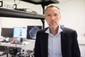
“Identifying tissues not only by their structure and morphology, as is done today, but also by their unique molecular signatures can assist in diagnosis and decision-making,” says lead author Alexander Golberg, professor of environment and earth sciences at Tel Aviv University. “We hope to rapidly find commercial partners to bring this method to the clinic.”