Available to watch now, Elekta explores quantitative MRI methods on MRI-guided radiotherapy systems
Want to learn more on this subject?
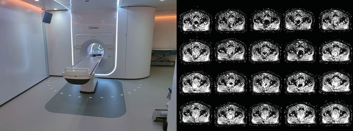
MRI-guided radiotherapy (MRIgRT) systems have the capacity to acquire functional, quantitative images in addition to the anatomical images required for online treatment guidance. This creates the potential to bring two important concepts in modern radiotherapy together: adaptive radiotherapy and biological targeting. Based on frequent anatomical and functional imaging, monitoring the changes that occur in volume, shape as well as biological characteristics, a treatment plan can be updated regularly to accommodate the observed treatment response. For this purpose, quantitative imaging biomarkers (QIB) need to be identified that show changes early during treatment and predict treatment outcome.
The first pilot studies have shown that QIB measurements are feasible on MRIgRT systems. However, the MR parts of these systems differ from regular diagnostic systems. This impacts the performance of QIB measurements and warrants technical validation. To ensure that the results of QIB studies on MRIgRT systems are also valid for diagnostic platforms outside the MRIgRT domain, QIB trials should be designed to establish reproducibility between systems. To identify if changes observed during the course of treatment are significant, the trials should include test-retest acquisition of the QIB prior to the first irradiation.
Within the Elekta MR-Linac Consortium, a working group on MRI biomarkers for response assessment is active in developing trial strategies, acquisition and analysis methods to make QIB research on Elekta Unity possible.
In this webinar, hosted by Prof. Uulke A van der Heide, an overview of the activities within the working group will be presented.
Want to learn more on this subject?
 Uulke van der Heide received his training as medical physicist at the department of radiotherapy of the University Medical Center in Utrecht, the Netherlands, and worked there as a medical physicist until 2011. Since then, he has worked as a medical physicist and senior group leader at the Netherlands Cancer Institute in Amsterdam, the Netherlands. He holds a chair as professor of imaging in radiotherapy at the Leiden University Medical Center.
His research group works on the improvement of target definition in radiotherapy by application of MRI and the development and validation of quantitative imaging methods for tumour characterization for radiotherapy dose painting. He further leads the MR-guided radiotherapy programme at the Netherlands Cancer Institute.