An electroretinogram (ERG) is a standard clinical method for measuring the function of the human retina. This procedure typically uses either a contact lens electrode or a fibre electrode to record retinal activity, both of which require physical contact with the eye, and therefore cause discomfort for the patient.
Researchers at Aarhus University in Denmark, led by Britta Westner and Sarang Dalal, have tested a potential replacement for these uncomfortable electrodes by using optically pumped magnetometers (OPMs), publishing their results in NeuroImage. OPMs are quantum sensors that detect magnetic fields that are over one billion times smaller than the earth’s magnetic field. Recent advances in quantum technology have enabled the design and commercial production of lightweight and flexible OPM sensors, such as those being incorporated into magnetoencephalography scanners.
While retinal response is typically detected via electrodes, which measure the electrical activity from the eye surface, the OPM sensors instead record the corresponding magnetic field induced by this electrical activity. The resulting magnetoretinograms circumvent the need for direct contact with the patient’s eye.
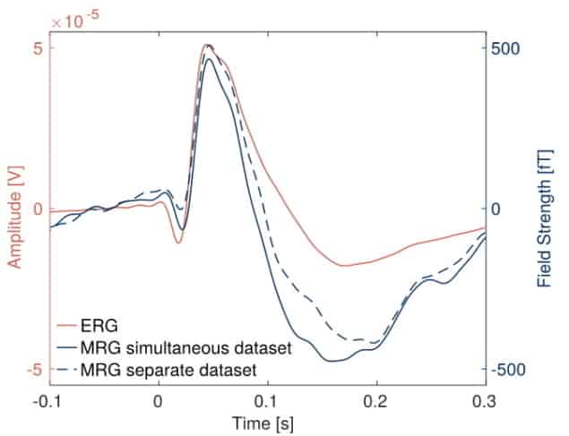
At Aarhus University, the researchers placed multiple OPMs close to a participant’s eye while performing routine retinal measurements, using a fibre electrode and a flashing light stimulation. During such a diagnostic test, a flash of light stimulates a negative potential in the retina, the “a-wave”, followed by a positive wave, the “b-wave”, which occurs at known times following the stimulus.

These features were clearly visible in the data recorded by the OPMs and subsequent comparison to the fibre electrode measurements showed a close match. To ensure the OPMs were not simply detecting signals from the electrode, the scientists repeated the experiment without the electrode on the eye and found very similar results.
Interestingly, artefacts due to eye blinks were reduced without the fibre electrode, as the OPMs offered a more comfortable scanning environment. However, the OPMs still suffered from a lower signal-to-noise ratio than the fibre electrode, likely due to a noisy magnetic environment.
As the magnetic fields originating from the retina are much smaller than the residual environmental magnetic field, the researchers operated the OPMs in a magnetically shielded room, to screen out some of this magnetic noise. They note that additional cost-effective active magnetic shielding could reduce this interference, resulting in a comparable signal-to-noise ratio to the ERG.

Quantum physics gives brain-sensing MEG scanners a boost
The team concludes that OPMs have the potential to replace fibre electrodes and contact lens electrodes to provide a contactless and comfortable clinical retinal scanning system. Furthermore, the flexibility of these magnetic field sensors will benefit neuroscientific research into human vision and the pathology of vision impairment.


