Available to watch now, Horiba Scientific explores the evidence that Raman microscopy is a key technique for chemical characterization
Want to learn more on the subject?
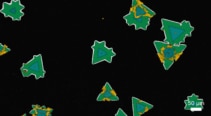 From materials to life or earth sciences, in many cases, solely obtaining the global chemical composition of a sample is not enough to characterize it completely. Spatial distribution and morphological information are also mandatory to give a full and realistic understanding of the sample studied.
Confocal Raman microscopy is the perfect technique to provide complete and deep chemical characterization of a sample. With our new LabRAM Soleil ultrafast imaging confocal microscope, the result of more than 50 years of HORIBA knowledge in spectroscopy, even for the most difficult sample, you can get easily and quickly a high-definition Raman image.
Join this webinar, presented by Thibault Brulé, to discover how LabRAM Soleil can solve your research challenges.
Want to learn more on the subject?
 Thibault Brulé is Raman application scientist at HORIBA France, working in the Demonstration Centre at the HORIBA Laboratory in Palaiseau. He is responsible for providing Raman spectroscopy applications support to key customers from various industries, as well as contributing to HORIBA’s application strategies. Prior to joining HORIBA in 2017, he conducted research on proteins in blood characterization based on dynamic surface enhanced Raman spectroscopy. He then applied this technique to cell-secretion monitoring. Thibault holds a MSc from the University of Technologies of Troyes, completed his PhD at the University of Burgundy and followed on with a postdoc fellowship at the University of Montreal.

