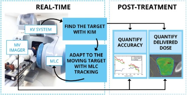
Real-time image-guided adaptive radiation therapy (IGART) is currently limited to expensive, dedicated cancer radiotherapy systems. The combination of two software programs that can be used with any standard linear accelerator – an image-based real-time tumour localization system and a real-time multileaf collimator (MLC) tracking system – offers the potential for widespread implementation of real-time IGART.
Researchers at the University of Sydney and the Royal North Shore Hospital in Australia have demonstrated the feasibility of performing real-time IGART with a standard linac by using two self-developed real-time systems: kilovoltage intrafraction monitoring (KIM) for measuring the position of a tumour during treatment; and an MLC tracking system. They have now used these combined software programs to treat eight prostate cancer patients participating in a clinical trial to evaluate KIM’s capabilities (Radiother. Oncol. doi: 10.1016/j.radonc.2018.01.001).
This clinical achievement represents another milestone towards affordable and widespread implementation of real-time IGART when stereotactic body radiotherapy (SBRT) is used to deliver radiation treatments. As an example, in Australia, there are currently only two very expensive dedicated linacs capable of real-time adaptation. An additional 200 standard linacs in the country could benefit from combined KIM/MLC systems, and potentially offer safer radiotherapy to many more cancer patients with tumours affected by body motion.
KIM is a real-time intrafraction tumour tracking system that provides six degrees-of-freedom (DoF) motion information during SBRT. Using the image-guidance system on a standard linac to acquire two dimensional (2D) projections of implanted fiducial markers, KIM reconstructs three-dimensional (3D) positions by maximum likelihood estimation of a 3D probability function, and makes additional computations to obtain 6 DoF motion information.
This capability enables real-time monitoring of a tumour’s position and rotation prior to and during SBRT. It tracks prostate motion in 3D with sub-millimetre accuracy, providing tumour targeting and enabling tighter treatment margins. KIM has been in development for over a decade, with research led by Paul Keall, professor of medical physics at the University of Sydney Medical School and director of the university’s ACRF Image X Institute.
KIM sends the tumour position signal in real-time to the MLC tracking code, which adjusts the leaf position to optimally align the treatment beam with the real-time target position. Real-time MLC tracking helps improve geometric accuracy. It also can be extended to correct for rotations and deformations observed in real-time.
Clinical trial
In the TROG 15.01 SPARK trial, researchers are testing the use of KIM to monitor prostate position during prostate radiotherapy delivered by standard linacs. Intrafraction motion can reduce the delivered radiation dose and increase the dose to organs-at-risk. The trial aims to measure the radiation dose of 48 patients who received SBRT with KIM, compared with the dose that would have been delivered had KIM not been used. The trial’s secondary outcomes include measuring KIM targeting accuracy, and quality-of-life, toxicity and biochemical cancer control for the participants.
Each patient received five 7.25 Gy fractions for a prescribed dose of 36.25 Gy to 95% of the planning target volume (PTV), delivered by a standard linac (Varian’s Trilogy) and MLC. Use of KIM for the total treatment generated an estimated additional kV dose of 0.4 Gy.
Thirty-nine out of 40 fractions were successfully completed using integrated KIM and MLC tracking, with one treatment using KIM and gating due to a technical reason. The geometric accuracy of the KIM system was excellent, with a mean accuracy within 0.2 ±0.6 mm in all directions. Prostate motion of between 3 and 7 mm occurred in more than half of the 40 fractions, but real-time IGART reproduced the planned dose more closely than when it was not applied.

The dose to the PTV and clinical target volume (CTV) were also consistently higher. An analysis of the treatment with the largest target motion, 8 mm throughout one fractional treatment, showed that the geometric accuracy of KIM was not affected by the magnitude or duration of this motion. In fact, 100% of the CTV received the prescribed dose with the use of real-time IGART, compared with 95% without it.
Keall told medicalphysicsweb that he is excited by the future opportunities that this achievement creates. “With the growing global awareness of the need for high-quality accessible radiotherapy, using the KIM and MLC software tools to bridge the technology gap between standard and high-end radiotherapy system capabilities can make real-time adaptive radiotherapy accessible to a much larger number of cancer patients than is currently possible,” he explained.


