Researchers in the US have developed nanosensors that can be directly inserted into a cell’s lipid membrane and be used to measure membrane potential. The devices, which are based on inorganic semiconductor nanoparticles, could potentially record action potentials from multiple neurons as well as electrical signals on the nanoscale – for example, across just one synapse.
Thanks to recent advances in inorganic colloidal synthesis, researchers can now make functional semiconductor nanoparticles whose size, shape and composition can be precisely controlled. Such nanoparticles can be used in applications as diverse as optoelectronics, biological imaging, sensing, catalysis and energy harvesting.
These nanomaterials can also be combined with biological cells to make highly sophisticated hybrid nanomaterials that outperform their purely biological counterparts. Until now, however, incorporating these particles into cell membranes has proved difficult. This is because they are often too big and have surface properties that can lead to non-specific binding on cell membranes. What is more, inserting nanoparticles into membrane bilayers is further complicated by the fact that their surfaces need to be functionalized so that the particles are inserted in the correct orientation.
Imparting membrane protein-like properties to nanoparticles
A team led by Shimon Weiss of the University of California, Los Angeles, says that imparting membrane protein-like properties to the nanoparticles might make it easier for them to target and insert into the lipid bilayer. This approach could be used to make membrane-embedded hybrid nanomaterials with useful functions. The researchers have now developed a way to do just this using rod-shaped nanoparticles and have also shown that the particles can be used to measure membrane potentials for the first time.
Weiss and colleagues employed a peptide-coating technique that they developed to make sure that the nanorods inserted themselves into the membranes in the correct direction – that is, perpendicular to the membrane surface. “This is important because rods inserted parallel to the membrane surface cannot detect membrane potentials across the membrane,” explains Weiss. “The coating technique itself involves adsorption of amphiphilic peptides with hydrophilic sequence segments aligning with the tips, and hydrophobic sequence segments aligning with the sides of the nanorods.”
Nanorods can sense membrane potential
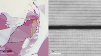
The researchers, reporting their work in Science Advances 2018;4:e1601453, confirmed that the nanorods inserted into cell membranes in the right direction by imaging them with transmission electronic microscopy.
Once inserted, these nanorods can sense membrane potential thanks to the quantum-confined Stark effect with single-particle sensitivity. “With further improvements, these nanosensors could potentially be used for simultaneous recording of action potentials from multiple neurons in a large field of view over a long duration and for recording electrical signals on the nanoscale, such as across one synapse,” say the researchers.
They add that they will now be improving the peptide coating and membrane-insertion process.