A novel non-invasive test for thyroid cancer works by asking the patient to sing at the same time. Developed by researchers in France, the ultrasound-based procedure could determine the health of a patient’s thyroid and help detect any cancerous nodules.
Thyroid nodules are common, but only a small percentage of such nodules are cancerous. Typically, fine needle aspiration is to detect malignant tumours, but only about 5% of thyroid cancers are detected in this way. As the majority of thyroid cancers are hard, the presence of cancerous tissue in the thyroid will increase its stiffness. This makes elastography – a technique that measures tissue stiffness – an ideal candidate for detecting cancerous nodules.
In this study, the researchers designed an experiment based on passive elastography, which extracts elasticity from the natural vibrations in living tissues. Specifically, they exploit the shear waves generated naturally by the human voice to measure the elasticity of thyroid tissue – a technique they call vocal passive elastography (V-PE). They report their findings in Applied Physics Letters.
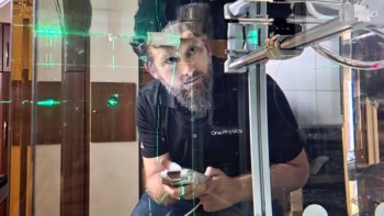
The researchers – from Université de Tours, CHU Dijon-Bourgogne and Université Bourgogne Franche-Comté – asked a volunteer to sing and maintain a monotonous tone at 150 Hz (roughly the frequency of the note D3), with a loudspeaker playing the same note to guide them. As the participant sings, vibrations in their trachea will induce shear waves in the surrounding thyroid gland.
The team tracked these waves using an ultrafast ultrasound probe placed horizontally against the surface of the neck. They then used correlation algorithms, based on time reversal methods initially employed in seismology, to compute the speed of the shear waves propagating through the thyroid.
If a tumour is present in the thyroid, the resulting increase in elasticity will cause the shear waves to accelerate. By superimposing a map of shear wave speed onto a thyroid ultrasound image, the researchers can mechanically characterize every point of the thyroid and find any abnormally stiff areas.
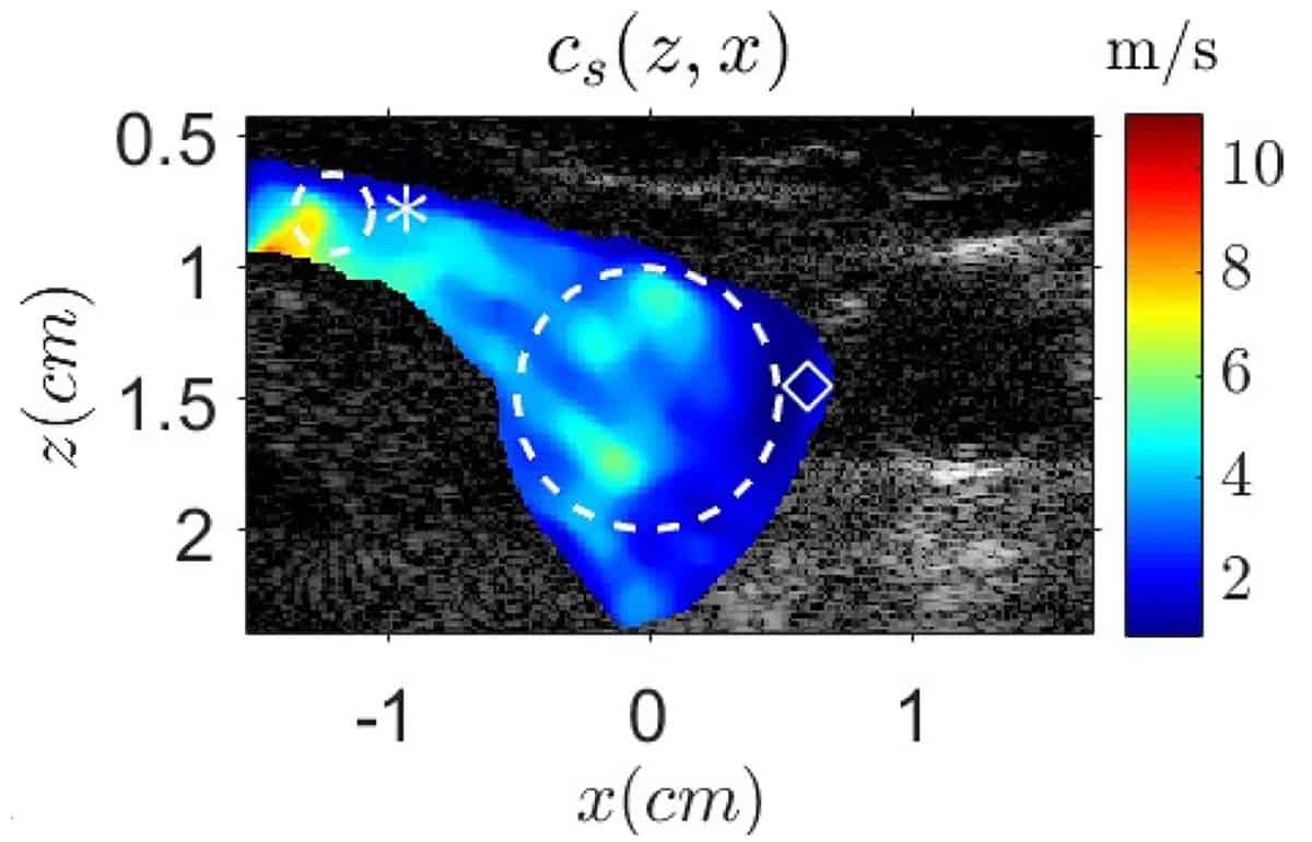
Using their V-PE method in a volunteer, the team measured the shear wave speed at every point within a mask surrounding the thyroid, with a pixel resolution of 150 × 150 μm. The mean shear wave speed was 3.2 m/s, ranging from 0.7 to 8.8 m/s. To validate their V-PE algorithm, they also used an AIXplorer ultrasound scanner to measure shear wave speed in the volunteer’s thyroid. The measured values agreed well with the V-PE results.
The researchers point out that V-PE is quick and easy to perform. It does not need any specialized equipment to be added to the ultrasound scanner and requires only about 1 s of data acquisition. The longest step is the data analysis, but they have developed a computer program to perform the required computations automatically. The team is now working to improve the user friendliness of the computer interface, as well as investigating the potential of V-PE in other areas near the vocal tract, such as the brain.
“Developing non-invasive methods would reduce the stress of patients during their medical exams,” says first author Steve Beuve in a press announcement. “Having to sing during a medical exam can perhaps help release some of the nervous tension even more.”