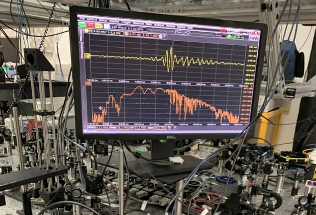
An infrared frequency comb small enough to fit on top of a table has been used to probe the structure and composition of a complex protein molecule. Scott Diddams at the National Institute of Standards and Technology (NIST) in Boulder, Colorado and an international team of collaborators built their system using a relatively simple laser setup, which can span the entire range of mid-infrared frequencies.
Large biological molecules such as proteins have incredibly complex and intricately folded structures, making them notoriously difficult to study. Typically composed of many thousands of atoms, proteins tend to vibrate and rotate at frequencies associated with mid-infrared light. Probing protein molecules with this light, and then measuring their characteristic absorption spectra, can yield important insights into their compositions, structures, and functionalities.
Such experiments have proven difficult because laser spectroscopy systems currently lack large enough bandwidths in the mid-infrared region to study the full range of protein resonances simultaneously. In addition, it is more difficult to accurately tune sources of mid-infrared light and to accurately detect it, than is the case for visible and near-infrared light.
Regular frequency intervals
Diddams’ team addressed these issues by probing protein molecules with a mid-infrared frequency comb generated using two phase-locked fibre lasers. This creates a series of short, bright pulses at regular frequency intervals across the entire range of mid-infrared frequencies – with the frequency spectrum of the light resembling the teeth of a comb. After interacting with the molecule, the light is detected by photodiodes detectors at a spectral resolution of 0.003 cm-1. Overall, the setup is small and simple enough to fit on a table top.
What is a frequency comb?
Diddams and colleagues tested their system on NIST’s monoclonal antibody reference protein. Composed of more than 20,000 atoms, this molecule is used to assess the quality of pharmaceutical treatments. By looking at the molecule’s absorption spectrum using the frequency comb, the team measured characteristic signatures called amide bands. These bands are used by biochemists to determine folding, unfolding and aggregation mechanisms in proteins. The researchers also detected sheet structures within the protein; verifying the results of previous studies that have suggested that chemical groups in the protein are connected in flat arrangements.
The team says that its frequency comb system could be combined with other techniques such as infrared atomic force microscopy to create a table-top system to determine the structure of proteins that would rival those at much larger synchrotron facilities. With further research, their techniques could also be used to store information within molecular vibrations and rotations – offering new technologies for quantum computing.
The research is described in Science Advances.


