A compact ultralow-field (ULF) brain MRI scanner that does not require magnetic or radiofrequency shielding and is acoustically quiet during scanning has been developed at the University of Hong Kong. The scanner’s low manufacturing and operating costs reinforce the potential of ULF MRI technology to meet the clinical needs of hospitals in low- and middle-income countries, as well as point-of-care medical facilities such as surgical suites and emergency rooms.
MRI is the most valuable clinical tool used for assessing brain injuries and disorders, but according to the Organisation for Economic Co-operation and Development (OECD), approximately 70% of the world’s population has little or no access to it. High-field superconducting MRI scanners (1.5 T and 3.0 T) are expensive. In addition to acquisition costs of around $1–3m, such scanners are costly to install due to infrastructure requirements, and have high maintenance costs. All of these factors represent a major roadblock in MRI accessibility.

MR imaging using ULF technologies offers the promise of accessible healthcare with scanners that are simple to onboard, maintain and operate. Led by Ed X Wu, Lam Woo Professor in the Laboratory of Biomedical Imaging and Signal Processing, the Hong Kong team developed a permanent magnet-based, low-cost, low-noise, low-power and shielding-free ULF brain MRI scanner.
The prototype system, described in Nature Communications, is based around a compact two-pole 0.055 T permanent samarium–cobalt (SmCo) magnet, with dimensions of 95.2 x 70.6 x 49.7 cm and a front opening of 29 x 70 cm for patient access. The scanner has a footprint of approximately 2 m2 and can be operated from a standard AC power outlet. The team estimates that the machine could be built in quantity with material costs under $20,000.
The scanner configuration allows the formation of images using various universally adopted protocols for clinical brain imaging, including fluid-attenuated inversion recovery (FLAIR)-like and diffusion-weighted imaging (DWI). By building upon the methodologies developed for high-field MRI scanners, the ULF system provides a high level of flexibility for the development of future ULF MRI protocols.
The researchers developed a deep-learning-driven electromagnetic interference (EMI) cancellation technique to model, predict and remove external and internal EMI signals from MRI signals. This EMI cancellation procedure eliminates the need for a traditional RF shielding cage. Meanwhile, the high temperature stability of SmCo removes the need for any magnet temperature regulation schemes to stabilize temperature-dependent fields.
Wu and colleagues optimized four of the most common clinical brain MRI protocols – T1-weighted, T2-weighted, FLAIR and DWI – to produce signal-to-noise (SNR) ratios and contrast characteristics similar to those of clinical high-field MR images.
After tests on phantoms, the researchers used the scanner to image 25 patients with neurological conditions (brain tumours, chronic stroke and chronic intracerebral haemorrhages), using these four protocols. The patients then underwent the same exams on the hospital’s 3 T scanner. Examinations averaged approximately 30 min with the 0.055 T scanner, compared with 20 min using the 3 T system.

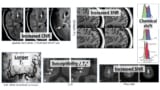
A senior clinical radiologist evaluated the patient scans to determine which specific lesions could be observed in the 0.055 T images. The prototype scanner detected most key pathologies in the exams of all 25 patients, with similar image quality to that produced by the 3 T scanner.
One major advantage of the new scanner is that it produces fewer artefacts when imaging implants such as metallic clips and cerebrovascular stents. “In using ULF, metal implants not only exhibit fewer artefacts, but also experience significantly less mechanical forces and RF-induced heating,” the researchers write. “The presence of paramagnetic (titanium and titanium alloys) and ferromagnetic (cobalt, nickel and associated alloys) materials in aneurysm clips and cerebrovascular stents did not induce gross artefacts.”
As such, the ULF scanner should enable MRI scanning of patients with metal medical implants or accident-related metal fragments, who would otherwise not be candidates for conventional high-field MRI.
Future opportunities
Wu believes that the ULF technology is not designed to compete with mainstream MRI, but to complement it. “With a field strength that is almost two orders of magnitude lower than the mainstream MRI, the image quality is inevitably less appealing simply due to the MR physics: lower field strength, weaker MR signal, less to play with,” he says. “However, MR signals and physics have many appealing properties at ultralow-field, in terms of data acquisition and image formation.”
“I believe that computing and big data will be an integral as well as inevitable part of future MRI technology,” Wu adds. “Given the inherently 3D, highly quantitative and ionization-free nature of MRI, I believe widely deployed MRI technologies will lead to immense opportunities in future data-driven MR image formation and diagnosis in healthcare. This will lead to low-cost, effective and more intelligent clinical MRI applications.”
High-field versus low-field MRI: is it time for a rethink?
The researchers chose to develop a ULF brain MRI due to “the immense need and value of MRI in the diagnosis and prognosis of various neurological diseases and injuries,” noting that roughly 30% of clinical MRI cases involve the brain.
Ultimately, they hope that the development of such ULF MRI technologies will enable patient-centric and site-agnostic MRI scanners to fulfil the unmet clinical needs across various global healthcare sites, with the potential to democratize MRI for low- and middle-income countries. To this end, the team is making the key code and designs they have developed freely available in a public online repository.