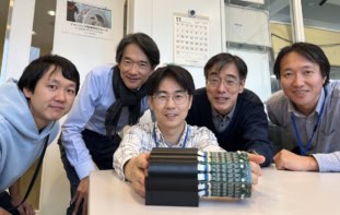
The multi-institutional EXPLORER Consortium is building the world’s first total-body PET scanner. The device, which has a sensitivity around 40 times higher than current clinical scanners, can capture a 3D image of the entire human body. And late last year, the EXPLORER PET/CT successfully produced its first human images.
Alongside, the consortium has developed two smaller-scale PET/CT scanners: the Mini-EXPLORERs. Mini-EXPLORER I is a long axial field-of-view (FOV) scanner containing detectors that are appropriate for clinical scanners. It is currently being employed to prototype total-body scanning in non-human primates.
“We wanted to build it for two main reasons — firstly, to learn more about issues that may arise with clinical-type PET scanners built with these longer geometries, and secondly, to develop new imaging methods with animals that could be translated for use in humans,” explains lead author Ramsey Badawi from UC Davis.
Mini-EXPLORER II, meanwhile, is designed to be a fully functional scanner for testing all the major components that are used in the human-scale total-body scanner. It also provides a high-resolution, high-sensitivity platform for clinical veterinary applications and human brain imaging. The team has now built a fully-functioning prototype Mini-EXPLORER II and performed the first in vivo imaging of a canine patient (Phys. Med. Biol. 10.1088/1361-6560/aafc6c).
Testing the design
Mini-EXPLORER II comprises a time-of-flight PET scanner with a ring diameter of 52 cm and an axial FOV of 48.3 cm, plus a 16-slice CT system. The PET scanner has two axial sections, each having 16 detector modules containing LYSO scintillator crystal arrays coupled to silicon photomultipliers.
Each section has a coincidence processor that receives single events from its own detector modules and can also share events with other coincidence processors — generating both “local” and “inter-section” coincidences. This design facilitates the integration of multiple PET sections to extend the scanner’s axial FOV.
“Mini-EXPLORER II uses the same PET detectors and electronics that are in the full-sized device,” Badawi notes. “The full-sized device has eight rings of detector modules and we needed to test that we could get the rings to talk to each other. The Mini-II has two rings of detector modules and uses the same communications board to get the rings to talk to each other.”
Badawi and colleagues performed a series of tests to assess the performance of the Mini-EXPLORER II. The prototype device exhibited an average system energy resolution of 11.7±1.5% and a timing resolution of 409±39 ps. Tests using the NEMA NU 2-2012 standard revealed a sensitivity of 52–54 kcps/MBq. The average spatial resolution at 10 mm from the centre of the FOV was 2.61 mm.
They also measured the peak noise-equivalent count rate, which was 314 kcps at 9.2 kBq/cc and 1712 kcps at 56 kBq/cc, using the NEMA NU 2-2012 and NU 4-2008 standards, respectively. The CT component of the system met all performance requirements for a diagnostic head CT imaging system.
Next, the researchers used various phantoms to evaluate image uniformity, spatial resolution and quality. Images of a long cylinder phantom that encompassed the entire axial FOV contained no significant artefacts. Scans of a mini-Derenzo hot-rod phantom clearly resolved the 2.4 mm rods, with 2.0 mm rods partially resolved. Reconstructed PET images of a Hoffman brain phantom showed the fine structures of the cerebral grooves and distinguished the white matter and ventricles.

In vivo study
Mini-EXPLORER II is an ideal size for veterinary applications, where the animals — typically dogs and cats — are too large for small-animal scanners and mostly too small for optimal imaging in clinical systems. The team scanned their first patient at the UC Davis School of Veterinary Medicine: a six year-old Corgi dog that had a small peripheral nerve sheath tumour surgically excised two years previously, followed by radiation therapy. The scan was performed to help characterize mild non-painful swelling.

PET images of the dog, recorded in three bed positions for 12 min each, exhibited excellent overall quality. Scans at the site of previous treatment revealed contrast uptake in the antebrachial musculature. Biopsy of this site confirmed a Grade I perivascular wall tumour, thought to have been radiation induced.
Badawi says that the Mini-EXPLORER II is now in routine clinical use for veterinary medicine. It is also being employed in clinical trials to develop new approaches for treating cancer in companion animals. “Some of these trials have direct relevance for future treatments for humans, so we are excited about that prospect,” he adds.
“One of the research projects that we are working on with the Mini-EXPLORER II, along with our colleagues in the School of Veterinary Medicine, is to develop methods for ultralow-activity imaging, specifically in the context of being able to track relatively small numbers of radiolabelled cells (for example stem cells) in the body,” co-author Simon Cherry told Physics World.


