
The development of a total-body PET scanner could revolutionize PET imaging, both for clinical applications and medical research. The EXPLORER project, a multi-institutional consortium, has now reported the successful performance of the mini-EXPLORER, a total-body, long axial field-of-view (FOV) PET scanner for animal imaging (J. Nucl. Med. doi: 10.2967/jnumed.117.200519).
The EXPLORER project was officially launched in October 2015 with a five-year $15.5m Transformative Research Award from the US National Institutes of Health (NIH) to researchers at UC Davis Biomedical Engineering. The award is funding the development of the world’s first high-sensitivity total-body PET scanner that can image the entire human body simultaneously. This capability could increase image sensitivity by a factor of 40, or reduce radiation dose by a factor of 40, compared with current clinical PET scanners. It could also decrease whole-body scanning time from 10-20 minutes to 15-30 seconds. A 40-fold increase in dynamic range could extend the imaging of radiotracers by more than five half-lives.
The UC Davis research team, jointly led by principal investigators Simon Cherry and Ramsey Badawi, has been conducting research on the development of a total-body PET scanner since 2005. Initial work involved optimizing the system design with computer simulations and better understanding its expected performance.
System performance
The mini-EXPLORER, now in use at the California National Primate Research Center, is a precursor to and a small-scale version of the human EXPLORER. The scanner was built using detectors and electronics from a clinical system (a Biograph mCT). It has 192 detectors arranged into eight rings for a ring diameter of 43.5 cm and an axial length of 45.7 cm.
The researchers used NEMA NU-2 and NU-4 phantoms to measure sensitivity and count rate performance. They investigated the reconstructed spatial resolution by imaging a radially stepped point source and a Derenzo phantom. They compared the performance of maximum acceptance angles between 14° and 46° to determine the effect of the wide acceptance angle.
The authors reported that the NU-2 total sensitivity was 5.0% and the peak noise-equivalent count rate measured with the NU-4 monkey scatter phantoms was 1741 kcps with the maximum 46° acceptance angle. The NU-4 scatter fraction was 16.5%, and the reconstructed spatial resolution was about 3.0 mm at the centre of the FOV. Both the scatter fraction and the spatial resolution showed little dependence on the maximum acceptance angle used (from 14° to 46°).
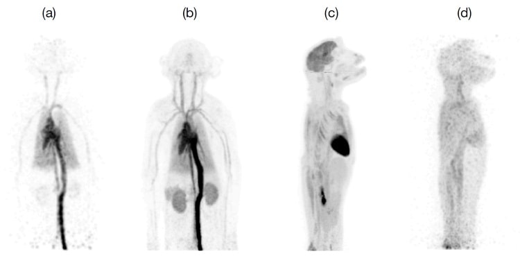
In vivo study
The team performed an 18F-FDG dynamic imaging study of a juvenile rhesus monkey with a single bed position. They verified that a wide acceptance angle can be used with a long axial FOV scanner to produce images with high sensitivity and overall excellent image quality. Early FDG distribution in major blood vessels and organs were visualized in one second frames shortly after injection. Reasonable image quality was obtained in a 40 minute scan at 18 hours post-injection, demonstrating the wide dynamic range of the mini-EXPLORER system and the ability to image very low activity concentrations.
Lead author Eric Berg, a postdoctoral researcher who received the College of Engineering’s award in 2017 for best doctoral dissertation on the development of the scanner, told medicalphysicsweb that the mini-EXPLORER was developed both to test some of the team’s hypotheses and to develop applications in non-human primates. He said that it is enabling the team to assess and study some of the effects of its much longer cylindrical geometry, to develop data correction and reconstruction algorithms, and to understand some of the features in the data expected to be encountered in the human EXPLORER scanner.
“The mini-EXPLORER is also allowing the team to develop applications in a relevant animal model that will ultimately be used in the human scanner,” Berg said. “These include studies of infection, inflammation and drug pharmacokinetics. We recently completed a study that demonstrated the distribution of 89Zr-labelled antibodies out to 30 days post-injection. We believe that this may be the longest time that a PET radiotracer has been successfully imaged after injection.”
Berg explained that the human EXPLORER scanner uses newer silicon photomultiplier-based detectors, as well as smaller scintillation crystals to achieve better spatial resolution. It will be much larger, with about 10 times more detectors and channels of electronics, with much greater, more sophisticated computing power and software for data acquisition and processing. The human scanner will also have an integrated CT for PET/CT studies.
The researchers are ahead of schedule to complete the human prototype scanner. “The underlying detector and electronics technology has been developed and tested, and it is operating at the required specifications,” said Cherry. “It has been incorporated in another small-scale prototype, the mini-EXPLORER II, which is being tested at the School of Veterinary Medicine. The first dog study was recently completed, and we have a good level of confidence in the scanner hardware. Remaining unknowns relate to whether the large volumes of data can be collected, sorted and reconstructed without problems. We also need to develop accurate data corrections for the many effects that can cause PET data to lose quantitative information if they are not properly handled.”


