

Pionium atoms arrive en masse

The strong interaction — which binds quarks together inside protons and neutrons — is described by a theory called quantum chromodynamics. Although the predictions of QCD have been confirmed for experiments with high energy transfer, the theory has not been tested as thoroughly at low energies.

A positive pion (π+) contains an up quark and a down antiquark held together by the strong force, whereas a negative pion (π–) contains the antimatter equivalent. Since pions have relatively low momenta in pionium, their interaction is described by low-energy QCD. The theory predicts that a pionium atom should decay into two neutral pions and have a lifetime of about 3 femtoseconds (3 x 10-15 seconds). In many ways pionium, which is denoted by A2π, is an exotic version of the hydrogen atom, although both its constituents have the same mass and both are unstable.

The first pionium atoms were created at the Serpukhov U-70 synchrotron in Russia in 1993. Although only about 270 atoms were detected, this result helped set a lower limit of 1.8 femtoseconds on the lifetime. In the DIRAC experiment, which includes physicists who were involved in the Serpukhov experiment, a 24 GeV proton beam from the Proton Synchrotron at CERN is directed onto a thin nickel target to produce pionium atoms in a proton-nuclear reaction.

The atoms then break up into positive and negative pions that are subsequently detected by a “double arm magnetic spectrometer” (see figure). This spectrometer has been specially designed to distinguish pairs of pions from pionium break up over the huge background of pairs produced in the same target.

The DIRAC collaboration was able to detect more than 5000 pionium break ups from a total of 6.4 x 10⁸ events, which will allow the lifetime to be determined to within statistical errors of 15%. The team hopes to reduce this error to just 10% in the future.

Angular pits boost DVD storage

DVDs store information in the form of simple, steep-sided pits each holding 1 bit of data. Although the storage capacity can be increased by writing the pits into different layers of the disk, it is still currently limited to around 4.7 Gb per layer. Now Peter Török and colleagues at Imperial College in the UK and co-workers at the University of Neuchâtel in Switzerland have realized that by giving the pits an angular sub-structure they could hold at least ten times more data.

To do this, the physicists needed to find a way of reading this angle rapidly, so as not to compromise the optical drive’s data rate. The solution was a combination of polarized light, a quadrant detector and light scattering analysis. The team has built a prototype using a 405-nm laser and the scheme, dubbed Multiplexed Optical Data Storage (MODS), is now being patented.

Although based initially on straight edge features, it turns out that the system also works with other pit geometries. “They do not have to be steps as long as they have a suitable asymmetry,” says Török. “The orientation is between 0 to 180°, but we can resolve 330 different orientations within the 0 to 180° angular range.”

Funded as part of the EU’s SLAM (Super Laser Array Memory) programme, the three-year project, which also involved scientists at Aristotle University of Thessaloniki in Greece, ended in May this year. Török believes that if his team can attract further funding, the first MODS disks, with a storage potential of around 250 Gb per layer, could be on the shelves between 2010 and 2015.

Despite having only a tenth of the storage capacity of MODS technology, it will be Sony’s BluRay that is the first to challenge DVDs’ domination of the audiovisual optical disk market. BluRay disks storing 25 Gb per layer, five times the capacity of current DVDs, are expected to be released towards the end of 2005 for the home market.

Table-top accelerators make progress

Conventional particle accelerators have to be hundreds of metres long to accelerate electrons to energies in the GeV range in, for example, synchrotron radiation sources. The machines used to accelerate particles to energies in the TeV for particle physics are even longer. Laser-produced plasmas are promising candidates for next-generation “table-top” particle accelerators because they can support electric fields that are thousands of times greater than those that can be produced in conventional accelerators.

The “laser wakefield” accelerator exploits the radiation pressure of an intense laser pulse to displace the electrons in a laser-produced plasma, leaving a large electric field in its wake. In 2002, Victor Malka of the Ecole Polytechnique in Paris (ENSTA) and colleagues showed that electrons could be accelerated to energies of 200 MeV over distances as short as a millimetre by “surfing” such a wakefield.

However, the beams produced in this and other experiments were of poor quality because the electrons had a wide range of energies. Now, three groups — including Malka’s team in Paris, a group led by Wim Leemans at the Lawrence Berkeley National Laboratory in the US, and a group led by Karl Krushelnick of Imperial College in London — have overcome this problem. By using variations on this approach the three teams have now produced monoenergetic beams.

Leemans and co-workers use pre-formed plasma channels to guide the laser beams over distances that are long compared to the natural diffraction distance of the laser beam. This diffraction would normally limit the distance over which particles surf the wake.

Krushelnick and colleagues employed a “forced” laser wake field approach, where the plasma wave actually “breaks” as the laser beam propagates through the plasma. This can lead to some of the electrons in the wave being “self-injected” into the wave. “It just so happens that when breaking first occurs, this bunch of electrons has a narrow energy spread, which is just what we want from our accelerator,” says Stuart Mangles from Imperial.

Malka’s group used their laser to create a “bubble” in the plasma, which traps and accelerates the electrons. “The main applications which will appear will probably be in radiobiology, medicine and chemistry,” says Malka. “In addition, our electron source will be perfectly suited for use in compact synchrotrons and free-electron lasers.”

All three groups now hope to reach GeV energies by accelerating the particles over longer distances.

The small world of Brazilian soccer

Roberto Onody and Paulo de Castro took data from a CD-ROM released by the Brazilian football magazine, Placar, and analyzed different networks formed by the 127 teams and the 13411 footballers who played in the Brazilian championships between 1971 and 2002. The most important results, says Onody, are that the professional careers of Brazilian footballers appear to be getting longer and, less surprisingly, that more Brazilian players are transferring to international clubs than ever before. There is also, he says, greater segregation between large and small clubs, often driven by television coverage of the game (Phys. Rev. E 70 037103).

Onody and de Castro found that the mean number of goals per game scored by a team was 1.03, while the average number of teams for which a footballer has played is 1.37. Moreover, the probability that a Brazilian footballer has played for a given number of clubs, or has played a given number of games, displays an exponential decay, whereas the probability that he has scored a certain number of goals follows a power law.

The probability that a footballer has played a certain number of games showed an unexpected critical value at 40 games. According to Onody and de Castro, this indicates that after a player has become famous, it is easier for him to continue playing football. It also appears that the distance between players — the so-called degree of separation or small-world effect — in the networks is very short at only 3.29.

In 2001 physicists at Warwick University in the UK analysed the number of goals scored in domestic football games in more than 150 countries, and found that the home team tends to win by an average of 0.51 goals per game.

Law-breaking liquid defies the rules

Solids usually melt when they are heated, and liquids turn into gas, although exceptions do exist when heating leads to chemical changes that cannot be reversed, such as polymerisation. However, a reversible transition in which a liquid becomes a solid when heated has never been observed until now.

Plazanet and colleagues prepared a liquid solution containing α-cyclodextrine (αCD), water and 4-methylpyridine (4MP). Cyclodextrines are cyclic structures containing hydroxyl end groups that can form hydrogen bonds with either the 4MP or water molecules.

At room temperature, up to 300 grams of αCD can be dissolved in a litre of 4MP. The resulting solution is homogenous and transparent, but it becomes a milky-white solid when heated. The temperature at which it becomes a solid falls as the concentration of αCD increases.

Neutron-scattering studies revealed that the solid phase is a “sol-gel” system in which the formation of hydrogen bonds between the αCD and the 4MP leads to an ordered, rigid structure. At lower temperatures, however, the hydrogen bonds tend to break and reform within the αCD, which results in the solution becoming a liquid again.

Molecular dynamics simulations by Plazanet and co-workers confirmed that the cyclodextrine ring becomes distorted as it is heated up to close to the solidification temperature. The hydrogen bonds within the αCD break and the hydroxyl groups rotate towards the outside, which allows a network of bonds to form between the different molecules. The team has found a number of cyclodextrine/pyridine systems that also become solid when heated, and is now looking more closely at the structure of the sol-gel system to understand the solidification mechanism in more detail.

Mars attacked by solar wind

Many theories have been put forward to explain how Mars changed from being a warm, wet planet to a cold, dry one. Recently it was estimated that a volume of water equivalent to a planet-wide ocean with a depth of between 14 and 34 metres could have escaped from the red planet during the past 3.5 billion years. Unlike the Earth, Mars does not have a magnetic shield to protect it from the solar wind, so particles from the Sun may have played a crucial role in shaping the Martian atmosphere.

Lundin and colleagues made in situ measurements of the solar wind flowing towards Mars, and the “planetary wind” flowing away from the planet. The planetary wind consists of volatile materials that are energised, ionised and accelerated by the solar wind as it penetrates Mars’ atmosphere.

They found that the solar wind can penetrate as deep as 270 kilometres above the Martian surface. Moreover, they found that positively charged hydrogen and oxygen ions flowing away from the planet can have energies as high as several keV at low altitudes, which means that they have enough energy to escape. According to Lundin and co-workers, the combined escape of hydrogen and oxygen ions might be evidence for a slow dehydration of Mars.

Explosive breakthrough

In the experiments a single photon has enough energy (75.5 electron volts) to knock both electrons out of a deuterium molecule, and the two nuclei then fly apart because they are both positively charged. By measuring the momenta of all four particles it is possible to learn more about what was happening inside the molecule.

Dörner and co-workers started by ionising a jet of deuterium molecules with polarised photons from the Advanced Light Source at the Lawrence Berkeley National Laboratory in the US. They used deuterium instead of ordinary hydrogen because it is heavier and therefore provides a higher target density for the photon beam. (A deuterium nucleus contains a proton and a neutron whereas a hydrogen nucleus contains just a proton.)

Next, they used electric and magnetic fields to accelerate the electrons created in the ionisation process in one direction, and the nuclei in another. The particles then left the region containing the electric field and drifted onto “micro-channel plate” detectors. For each particle, Dörner and co-workers were able to measure how long it took to reach the detector and the position of impact on the plate. This allowed them to calculate the initial momentum of all four particles and build up a 3D image of the photo-fragmentation “explosion” (see figure).

The results show that the behaviour of the electrons is strongly influenced by the separation of the nuclei at the instant the photon is absorbed. The experiment could lead to a better understanding of many physical and chemical processes through improved knowledge of the quantum dynamics of many-particle systems.

Radioactivity speeds up

Beryllium-7 can decay into lithium-7 through a process in which the beryllium nucleus captures one of its own electrons. This electron is then absorbed into the nucleus, where it combines with a proton to form a neutron. Increasing the density of electrons surrounding the nucleus can increase electron-capture decay rates because more electrons are likely to be captured and absorbed. The electron density can be increased by external factors, such as chemical environment or pressure.

Tsutomu Ohtsuki and co-workers at Tohoku University and Yokohama National University began by inserting beryllium-7 atoms into carbon-60 cages using a nuclear recoil implantation technique. Next, they measured the decay rate of the encapsulated beryllium — denoted 7Be@C60 — with a gamma-ray detector and found that its half-life was 52.68 days. This was 0.83% shorter than the half-life they measured for pure beryllium (53.12 days). The half-life of a radioisotope is defined as the time it takes for half of the original amount of material to decay.

The team says that the faster decay rate can be explained by the fact that the dense cloud of electrons in the carbon-60 cage increases the electron density at the nucleus. Moreover, the particular orbits of these electrons further increase the electron density at the nucleus.

Increasing the decay rate by just 0.83% will have little effect on radioisotopes with half-lives of thousands or millions of years. However, Ohtsuki and co-workers say that their results will help identify the sort of environments that increase the decay rate. These could include the high-pressure conditions found inside neutron stars.

Microscope focuses on sub-Angstrom scales

Sub-Angstrom imaging has been a long-standing goal for electron microscopists because it would allow structures to be studied at the level of single atoms. Although sub-Angstrom information can be obtained by post-processing electron micrographs, it has not been possible until now to obtain this information directly.

A scanning transmission electron microscope builds up an image by scanning an electron beam across a sample and measuring the parts of the beam that are transmitted back from the sample. However, the aberration — or blurring — that is caused by the magnetic lenses that are used to focus the electron beams has limited the resolution of these instruments to around 1.5 Angstroms (1.5×10⁻¹⁰ metres), which is slightly larger than the typical distance between atoms.

The resolution of an electron microscope increases as its aperture becomes larger but in the past lens aberrations have caused the images to blur once the aperture has reached a certain size. To overcome this problem, Stephen Pennycook of Oak Ridge and colleagues fitted an aberration corrector, made by Nion, onto their scanning transmission electron microscope. This corrector is a lens that employs software that is capable of analysing all axial aberrations in the microscope in less than a minute, and then making automatic adjustments to compensate.

The Oak Ridge-Nion team tested its device by imaging a silicon crystal in which the columns of atoms are known to be 0.78 Angstroms apart. Before correction, the optimum resolution for images was 1.3 Angstroms, but after correction it was possible to resolve the individual columns of atoms (see figure). The technique allowed the team to effectively double the aperture size of the lens.

“You can think of aberration correction as a pair of spectacles for a microscope,” says Pennycook. “Previously the vision was blurred but now we can see twice as clearly as before.”

“Seeing atoms more clearly allows us to see materials better, to understand how atoms go together so we can understand how things behave the way they do,” he adds. “The advance benefits a vast array of fields, from chemical sciences, material science and nanotechnology — everywhere people want to see what they have made.”

The team now plans to explore the possibility of using its device to image in 3D.

Nanotubes feel the force

Nanoelectromechanical systems or NEMS could be used for a wide variety of applications, including ultra-sensitive mass and force detection. In these devices a mechanical element moves in response to an external force, and a highly sensitive detector is used to record the displacement of the mechanical element.

Carbon nanotubes — rolled up sheets of graphite just nanometres in diameter — could be ideal candidates for making such devices because they are able to withstand large tensile stresses. This means that they could operate over a wide range of high frequencies, as is needed for quantum mechanical applications. Moreover, a nanotube can act as a transistor and is therefore capable of sensing its own displacement. This allows it to be used an electronic detector.

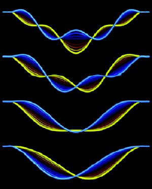
Paul McEuen and colleagues made their device by suspending a nanotube over a trench — typically 1.2 to 1.5 microns wide and 500 nanometres deep — between source and drain electrodes made of gold. The gate electrode lies underneath the nanotube, which itself is effectively clamped at both ends (figure 1).

The Cornell physicists adjusted the tension in the nanotube by varying the voltage at the gate electrode. This also had the effect of vibrating the nanotube. They then detected this vibrational motion by measuring the change in conductance of the nanotube as it moved through the electrical field of the gate electrode.

McEuen and co-workers showed that they could tune and measure the resonance of the nanotube over a wide range of frequencies — from 3 to 200 megahertz — by simply varying the gate voltage (figure 2). They were able to detect displacements as small as 0.5 nanometres with a force sensitivity that was within a factor of ten of the best values measured at room temperature to date.

Copyright © 2026 by IOP Publishing Ltd and individual contributors