

New features will be added to PhysicsWeb over the next few months to enhance the service offered to users.
New features will be added to PhysicsWeb over the next few months to enhance the service offered to users.
Future archaeologists who come across books from our era would, he says, find it difficult to tell the difference between ‘new age’ science books full of “junk science” or those containing scientific fact. This is why Lovelock believes that a well-written record of scientific knowledge designed for an intelligent reader is needed. The book should be clear, unbiased and constantly updated. It would explain the theory of natural selection and the principles of medicine, engineering and thermodynamics. It would also cover what we know about the universe. And to guard against future obsolescence, Lovelock argues that such a “survival manual” should be written on paper, not stored on computer.
The middle of Antarctica is an ideal place to look for ‘survivors’ of the burn-up because material in the region suffer little, if any, chemical or physical weathering and there is very little contamination. The snow and ice also increase the chances of finding extraterrestrial material. Taylor and colleagues realized that micrometeorites would concentrate at the bottom of the well. Moreover, as ice core samples taken around the base have accurately aged the ice layers, they could also estimate the age of material trapped in the ice.
The team built a remote-controlled robot to vacuum material off the well bottom – which is 20 meters underwater and 100 meters below the ice – without contaminating the water supply. The team estimated that filters attached to the robot collected 99% of the particles with diameters between 50 and 2000 microns – including 0.2 grams of material of exterrestrial origin. This is the largest collection of earth-based micrometeorites ever collected. Comparisons with data from experiments in Greenland and the space-based Long Duration Exposure Facility showed that 10% (±2%) of the particles in the range 50-700 microns reach the Earth’s surface.
Neutrinos travel at the speed of light and only interact very weakly with matter, which makes them very difficult to detect. This is why neutrino detectors need to be large and built underground – otherwise cosmic ray interactions will mimic the neutrino signal. When a neutrino does interact with the heavy water in the tank, a flash of Cerenkov radiation is emitted and recorded by some of the 10000 photomultiplier tubes that surround the tank.
Astronomers hope that the observatory will help solve the solar neutrino problem – the fact that existing experiments detect less than half the flux of neutrinos from the Sun predicted by theory. One possible explanation is that electron neutrinos are oscillating into muon neutrinos that cannot be detected by the other experiments – but this would require neutrinos to have a non-zero mass, which would have enormous implications for particle physics and cosmology. The observatory should also be able to detect neutrinos from other astronomical events such as supernova explosions.
The opening ceremony was broadcast over the Internet and included remarks from Stephen Hawking and two Nobel prize-winners – Bertram Brockhouse and Richard Taylor.
Superconductors lose their unique properties above a critical current density of about 105 A cm-2. The best candidates for high-current applications – silver sheathed tapes of the bismuth-lead-strontium-calcium-copper-oxygen alloy known as BSCCO – reach about 25% of this value. To explore the factors limiting current performance, the researchers analyzed some of the best multi-filament tapes available. They found that individual filaments had critical current densities much higher than the average value of the tape.
Electron micrographs and magneto-optic images of the tapes show a series of cracks which reduce the critical current density. The researchers also found that currents along the crystalline planes of the material were sometimes blocked by a series of nanometre and micrometre scale barriers.
The team point out that although cracks are very difficult to engineer out of the production process, it should now be possible to design new processes to eliminate them.
The sum is substantial – domestic spending on basic research is about IR£2m each year; the country also receives IR£20m from the EU. “If the money is not released very soon, there will be one very significant row, ” says Vincent McBrierty, a polymer physicist at Trinity College Dublin.
The Office of Science and Technology launched its usual call for proposals in December. “The applications were assessed, ranked and graded, then at the last minute, the plug was pulled, ” says John Donovan of the Irish Research Scientists Association. “The people losing out are those with unfunded projects, ” he says. The new work was due to start in the autumn.
The Office of Science and Technology is now hoping that the new projects will be funded by the Department of Education and Science. “We are in discussion with the department about whether they will use their money for our research start-ups this year, ” says Michael Fahy, who was head of the Office of Science and Technology until the end of last month. “[The issue] needs to be resolved…it should have been done yesterday, ” he adds.
The Foresight programme was launched in 1994 to improve wealth creation and the quality of life in the UK. It has been led by 16 panels of business executives, scientists and government officials, who have tried to identify and respond to future market trends and technological opportunities. The panels cover most sectors of the economy, from IT and energy to transport and defence.
The government thinks that the Foresight panels should remain at the heart of the programme, but it may change the number and remit of the panels. It also wants the panels to enrol a wider range of members, interact more closely with each other, and concentrate more on the global market. It is also keen to add new themes such as sustainable development, the ageing population and crime control. The new panels will be appointed in November.
“Foresight has started a very important process, ” says John Taylor, director of Hewlett-Packard Laboratories in Bristol and chairman of the IT, electronics and communications (ITEC) panel. “It has allowed industrialists and academics to get together and identify the fertile areas for investment, ” he says. “It has not been about picking winners, but it has helped us to look ahead dispassionately at what needs to be done.” Taylor adds that the ITEC panel must break away from talking to those academics and industrialists who already know about the benefits of IT and speak to people who are starting up new firms or designing new products.
In the last several years, my team at the University of Utah has developed a new seeding technique that can clear supercooled cloud and fog on a scale several times larger than a commercial airport. The method, which can be applied from a vehicle on the ground, has been used to clear airports, cities and roads, and has recently helped NATO peace-keeping operations at Bosnian airports.
The technique works with supercooled clouds and fogs, which consist of small water droplets at temperatures below freezing. Such fogs often form when the natural mechanism for ice formation is ineffective, and they often occur in mountain valleys and plains during cold seasons. In this situation, the supercooled droplets can be transformed into snow by introducing “seed crystals” into the fog.
Seeding causes a large number of small supercooled droplets at high vapour pressure to evaporate and recondense onto a smaller number of seed crystals at a lower vapour pressure. When the seeding works correctly, the grown crystals will fall out as snow and rain, leaving a clear space behind. For the technique to work, the seed material must cause large numbers of ice crystals to form.
Research into cloud seeding began in 1946, when Vincent Schaefer of General Electric noticed that a small piece of solid carbon dioxide (dry ice) can generate a large number of ice crystals in a laboratory-generated supercooled fog. He then went up in a small aircraft and dropped about 1.5 kg of crushed dry ice into supercooled stratocumulus clouds over western Massachusetts, and found that snow crystals did indeed fall out from the cloud. In the following year Bernard Vonnegut of General Electric found that particles of silver iodide can also generate large numbers of ice crystals if the cloud is cold enough.
With these reports, the world started to anticipate the day when people would be able to control the weather. In reality, however, a statistical approach was taken to investigate the technique further – much like the evaluation of a new medicine – which meant that there was no real investigation of the cause and effect of cloud seeding. The conditions needed to be fixed to prove statistically that seeding was effective, but the conditions should be varied to find the optimum method. Projects often proved a useless result, or failed to produce an effect that was physically convincing.
This problem was recognized in the 1970s. Emphasis then shifted to a scientific study of cloud processes, but the engineering of the seeding technique remained unexplored. At Utah we have focused on clarifying the fundamental processes involved in cloud seeding and on developing a practical seeding technique that makes best use of the feedback mechanisms involved.
This work has shown that the seeding reaction begins with ice formation or nucleation, and that this can occur through two possible mechanisms. In homogeneous nucleation, typically observed when seeding with dry ice, strong cooling of sublimating dry-ice pellets forces water vapour to condense in the surrounding air and the droplets freeze immediately. Heterogeneous nucleation is more complicated, and requires the help of foreign particles such as silver iodide.
Today, silver iodide is the seeding agent of choice because it can be released in dry or warm air at low altitudes, while dry ice pellets have to be dropped from high altitudes. Silver iodide also produces more ice crystals at lower temperatures. I have also observed the same behaviour with organic ice nucleants such as metaldehyde.
Experiments have shown that seeding with dry ice and liquid carbon dioxide can lead to ice nucleation that is almost independent of temperature. This is an advantage when forming ice crystals, since the heat generated in the phase change from supercooled liquid to solid causes the air containing the crystals to rise, and the maximum growth rate is sustained because the number of crystals produced does not depend on temperature.
An effective way to convert a large cloud mass to snow and rain is to ensure that the seeded particles that rise to the top of the cloud on a thermal fall back into the cloud. For this to happen the crystals must grow, which in turn suggests that they must co-exist with the supercooled droplets for a significant amount of time.
Since the supercooled droplets disappear as the crystals grow, more of the supercooled cloud is entrained into the thermal through eddies generated at its base. In the case of dry ice or silver iodide, a vertical ice plume is generated: the buoyant energy associated with the thermal is converted into an accelerating updraft with a low resistance force, similar to cigarette smoke. However, the crystal growth does not last long enough for crystals to fall back through the rising ice plume, and so this technique can only treat small volumes.
In contrast, the technique that we have developed ensures that the thermal lies horizontally. In this case the buoyant energy is converted effectively into turbulent energy, which means that the rising thermal has a slow updraft velocity. To maximize the interaction time of the rising thermal with the supercooled cloud, the seed crystals are generated at the bottom of the supercooled zone, just above the 0 °C isotherm. The seeding then induces a slow but large dynamic effect that lifts the underlying layer of moist air and increases the cloud volume that can be modified. It therefore makes most effective use of the energy created in the phase change.
Using this technique, we have sprayed liquid carbon dioxide – a homogeneous ice nucleant – directly into supercooled stratus clouds over the Great Salt Lake. This generated an optical effect known as undersun and then completely cleared the cloud over an area a few kilometres wide.
We have also used ground-based vehicles to seed supercooled fogs at night. This leads to the formation of light “pillars” – vertical streaks of light – above and below street lights and other light sources, which indicates that the ice crystals are orientated horizontally. The seeding cleared areas of up 100 km2 and caused about 1 cm of snow to fall on the ground.
Seeding of supercooled fog early in the morning often leads to other optical effects called parhelia, which are mock images of the Sun again produced by horizontally orientated ice crystals. The liquid carbon dioxide used in the seeding is either an industrial by-product or comes from natural gas, and does not alter the amount of carbon dioxide in the atmosphere.
The technique is already being used to clear supercooled cloud and fog, and it is most effective in targeting the effect at a particular region. For cloud seeding, it is more effective to inject the seed crystals from the air, and we anticipate that radar will be used to guide unmanned aircraft to the correct seeding position. Fog can be cleared from the ground, and it should be possible to develop a simple device that could be used to clear fog as and when it occurs.
One side effect of the increasing emphasis on maximizing the economic return on public investments in science and technology has been the growth of “foresight exercises” around the world. Pioneered by the Japanese government in the early 1970s, foresight has mushroomed in the 1990s. The basic idea is to identify the likely social, economic and market trends in all sectors of a nation’s economy over the coming 10-20 years, and then identify the developments in science and technology required to address future needs. The normal approach is to divide the economy into 15 or so sectors and set up a panel of experts from business, industry, government, universities and elsewhere to cover each one. These sectors are pretty similar across the globe: IT and electronics, materials, health and medicine, the environment, transport, agriculture and so on.
It is difficult to judge how successful any of the exercises have been, and it will be just as difficult to do so in the future. However, one important lesson has been learnt so far: foresight is not an end in itself and it is essential to make the results as widely known as possible. This is one of the themes to emerge from The Future in Focus: a Summary of National Foresight Programmes, a collection of short papers published by the UK Office of Science and Technology.
Japan is the one country where we can try to gauge the success of foresight. Since 1971 the Japanese government has published a major Technology Forecast Survey every five years or so. An analysis of the first survey has shown that it is easier to predict the future in some areas than in others. More than two-thirds of the predictions made about telecommunications and agriculture, forestry and fisheries were correct, whereas only one-quarter of those about energy and life sciences came true. However, many experts felt that it was more important to make management and policy-makers aware of gradually occurring trends in science and technology than it was to make exact predictions about the future. The creation of new contacts and networks was also seen as a useful spin-off from foresight.
A common problem is how to make the best use of the vast amount of information that is gathered. The results of a “Delphi” foresight survey carried out in Germany in 1993 were influential in setting public spending priorities, but further dissemination was limited. Greater emphasis will be placed on communicating the results of the second exercise, which was completed in March, to small- and medium-sized enterprises. Indeed public relations agencies have already been invited to tender for this work.
The situation in the UK is similar. The results of a major foresight programme were published in 1995, and by the start of 1997 over £350m of private and public sector funds had been committed to initiatives reflecting foresight priorities, although little of this was “new” money. However, programme officials admit that the findings of the survey were not used to their full potential. The UK is about to start on another round of foresight.
Surprisingly, perhaps, the US does not use foresight, although it does run similar exercises with varying degrees of success. The two-yearly Critical Technologies report, for example, is said to lack influence because the steering group that prepares it is dominated by academics. On the other hand, technology road maps prepared by US industry groups in areas such as semiconductors and optoelectronics have been more successful because they have focused on what a particular industry must do to survive and prosper in an era of increasing competition and change.
One result of foresight is that it will no longer be possible to rely on past glories (or anecdotes about Michael Faraday) when trying to argue that a particular line of research will eventually lead to applications. A major challenge is to relate the work of the panels, which focus on market sectors, to a nation’s research base that will largely be organized along completely different lines. For this reason physicists and the organizations that represent them would be well advised to exert as much influence as possible on the various foresight activities now underway, and to interpret and exploit the reports in the widest and most creative manner possible.
It is impossible to overstate the importance of proteins to plant and animal life. Much of the tissue in the human body is made of protein, as are all of the enzymes that catalyse reactions in the body, the globins that transport and store oxygen, the antibodies responsible for the body’s immune response and hormones such as insulin.
Proteins are macromolecules made from combinations of the 20 naturally occurring amino acids. A typical protein contains about 300 amino acids, although some proteins can contain as many as 1000, and multi-macromolecular protein and nucleic-acid complexes represent further levels of complexity. Amino acids are small molecules that are made up mostly of carbon, hydrogen, nitrogen and oxygen, although two amino acids also contain sulphur.
The order of the amino acids in a protein is determined by the sequence of “base pairs” in the deoxyribonucleic acid (DNA) that is found inside every living cell. In humans this DNA is divided among 23 pairs of chromosomes that collectively make up what is known as the human genome. A gene is the sequence of base pairs that carries the code for a single protein and there are about 100,000 genes in the human genome. Scientists from all over the world have joined forces to determine the order of the base pairs in the human genome and physics-based techniques are playing their part in that effort (see “Physics and the Human Genome Project” by Norman Dovichi in Physics World September 1997).
Just as important as chemical composition, however, is the shape or conformation of the protein because this determines its detailed chemical and biological function – be that enzyme catalysis, viral infection, oxygen transport or the immune response. The shape is determined by the orientation of each amino acid relative to its two neighbours, with the amino acids linking in a way that is energetically “comfortable”. Although there are only three or so sterically acceptable orientations for each pair of amino acids, the total number of shapes possible for a typical protein containing 300 amino acids is thereby huge, about 3300.
One of the great mysteries of biology is how a long chain of amino acids “folds” into its final working (i.e. active) shape within seconds – if the protein followed a random walk to its final structure, it would take much longer. This is a problem that has attracted the attention of many physicists as it has parallels with systems studied in statistical mechanics.
Progress has been made in terms of the secondary structures within a protein: two commonly found substructures are the so-called alpha helices and beta sheets. However, large parts of the structure cannot be predicted. Moreover, even for those substructures like helices that can be predicted, the precise placement of the atoms or chemical groups that are central to the protein’s function cannot. These side chains determine the reactivity and intermolecular “recognition” of the proteins.
There is therefore great demand for fast and efficient techniques to determine the 3-D structures of proteins in terms of bond distances and angles. Many physics-based techniques have been developed for this purpose. Nuclear magnetic resonance, for example, does not require the protein to be available in a crystal form, but is restricted to small proteins. Electron microscopy is used when only 2-D arrays of proteins are available, for example for membrane-bound proteins, which are generally difficult to crystallize. Some 40% of protein structures in a genome are membrane bound.
The most precise technique, however, and the technique capable of tackling the largest molecules is X-ray crystallography. Moreover, around 60% of proteins (being non-membrane bound) are very amenable to crystallography. This technique, which has a long history, has recently been revolutionized by the development of synchrotron radiation sources operating at X-ray wavelengths. Researchers have also made progress in overcoming the two principal bottlenecks in this quest: the growth of high-quality crystals and the well known crystallographic-phase problem.
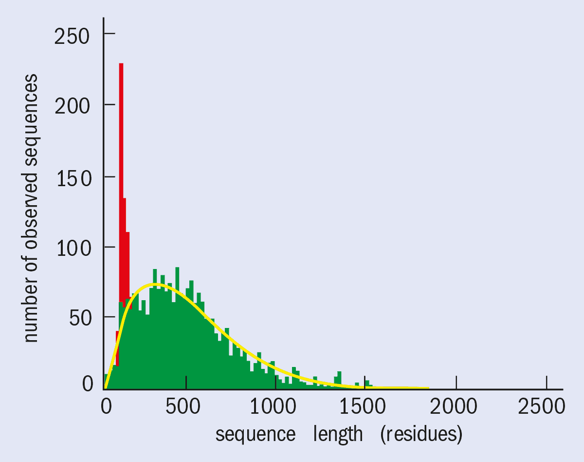
To put the challenge in perspective, it should be noted that there are about 100,000 proteins in the human genome. The yeast genome, which comprises about 10,000 proteins and has already been sequenced, tells us more about the technical challenges. A typical protein contains about 2300 non-hydrogen atoms (i.e. 300 amino-acid residues), but some proteins contain as many as 7000 non-hydrogen atoms (figure 1). The proteins in the human genome are expected to follow a similar distribution.
To date there are some 7000 protein structures in the Protein Data Bank held at the Brookhaven National Laboratory in the US, and this number is doubling approximately every two years, although not all of these structures are “new” proteins since variants (e.g. mutants) of a given protein structure are also deposited. Some 1400 of the 7000 structures are human and 6000 have been determined by X-ray diffraction.
On the basis of current achievements, the determination of the 3-D structures of many proteins in whole genomes has become a realistic prospect. Such a goal is being discussed at the highest levels in synchrotron radiation facilities around the world as part of a possible Genome 3D Structure Determination Project.
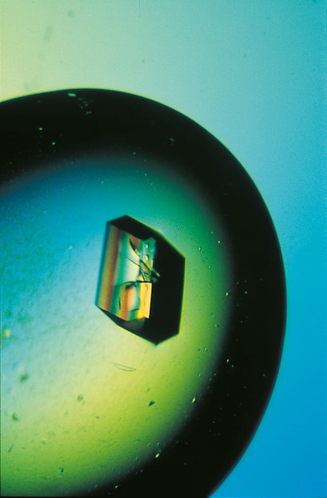
Protein crystals are quite different to the crystals studied by most physicists. A protein crystal typically has dimensions of ~ 0.5 mm and contains about 1015 protein molecules in a periodic array. However, protein crystallographers are primarily interested in the arrangement of the atoms inside the protein molecule itself, rather than the arrangement of the molecules inside the crystal.
Around half of the protein crystal is actually made of liquid: indeed, a protein crystal becomes disordered if it is allowed to dry out. A small fraction of the solvent binds to the protein to form an ordered shell (or shells), but most of it is found in “bulk solvent channels”. These channels allow the diffusion of smaller molecules or “ligands” into the crystal (e.g. molecules whose reactions are catalysed by enzymes or, just as important, molecules that inhibit enzyme action). These protein-ligand complexes can also be studied by X-ray crystallography, and this sort of work is increasingly being used in the pharmaceutical industry for rational drug design.
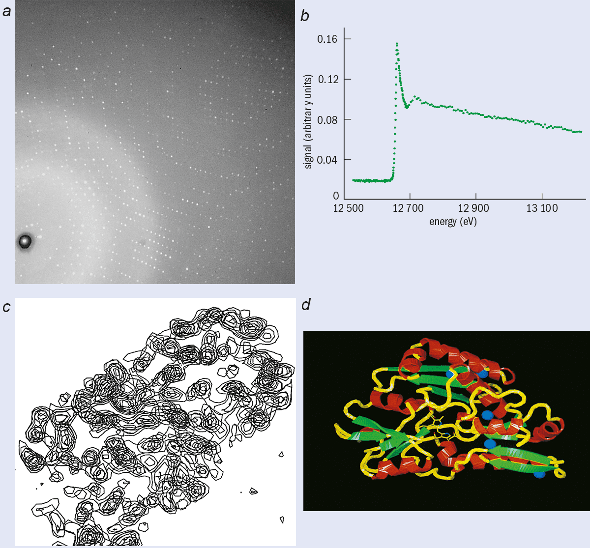
X-ray crystallography relies on the scattering of X-rays by the periodic arrays of atoms and molecules that comprise a single crystal. The scattered X-rays interfere to produce a diffraction pattern that contains a large number of spots in regular positions (see right). Each spot or reflection is governed by Bragg’s law l = 2d sinq, where l is the X-ray wavelength, d is an interplanar spacing in the crystal and 2q is the scattering angle with respect to the incident X-ray beam. For a crystal continuously rotated in an X-ray beam of one particular wavelength, spots light up as the crystal momentarily reflects and then disappear. A complete set of diffraction spots can be measured for a full 360° rotation of the crystal. In practice it is found that the pattern often repeats itself after a smaller rotation (e.g. 90°) due to the internal symmetries of the crystal.
The first crystal structure to be resolved was sodium chloride (NaCl) in 1913, an achievement for which Sir Lawrence Bragg shared the 1915 Nobel Prize for Physics with his father Sir William Henry Bragg. Lawrence Bragg deduced the structure of NaCl by comparing the diffraction patterns of crystals with similar compositions, particularly sodium chloride and potassium chloride (KCl). Although many of the spots were of similar intensity, some of those in the NaCl pattern were missing in the KCl pattern.
Bragg relied on the fact that both crystals were actually diffraction gratings and that the X-rays would therefore undergo constructive or destructive interference from the atomic electrons, producing strong and weak spots, respectively. In particular, Bragg realized that since the potassium and chloride ions both had 18 electrons, destructive interference would lead to missing spots if the crystal contained an alternating array of potassium and chlorine ions in three dimensions. Sodium chloride had a similar structure.
This intuitive approach has been replaced by methods based on Fourier transforms (figure 2). To reconstruct the 3-D arrangement of atoms and molecules responsible for the diffraction pattern with this technique we need to know the amplitude and phase (relative to the incident beam) of each spot in the pattern (see box “Multi-wavelength crystallography”). However, a basic problem is that although the intensity of each spot can be measured, the precise phase cannot. This is a technical rather than fundamental limitation. The problem is that radiation with extremely short wavelengths (~ 1 Å) is needed to probe the interatomic distances (also a few Å) and it is extremely difficult to measure phase at such short wavelengths.
The electron density, ρ(x,y,z), of the unit cell in a crystal is related to the diffraction data by a Fourier series
where ρ is in units of electron charge per Å3, V is the volume of the unit cell, h, k and l are integers used to label the diffraction spots in three dimensions, |Fhkl| is the amplitude of the hkl-th spot and αhkl is its phase angle. As explained in the text, the big challenge in X-ray crystallography is to determine these phase angles, with a variety of methods having been developed for molecules of different sizes.
The traditional method for doing this for protein crystals has been multiple isomorphous replacement. In this technique, diffraction patterns are collected for the native protein crystal and for two derivative protein crystals in which some atoms have been chemically replaced by heavier atoms. However it can be difficult, and sometimes impossible, to find suitable heavy-atom replacements, since the replacement atom must not disturb the crystal lattice or protein structure. It is also labour intensive.
With modern multi-wavelength anomalous dispersion (MAD) techniques, however, it is possible to introduce a metal atom such as selenium or xenon directly into the protein and to exploit changes in the scattering properties of the atom as a function of synchrotron radiation wavelength. This means that fewer crystals are needed – only one when selenium is used and only two for xenon.
MAD methods rely on the changes to diffraction spots caused by the metal atom. For this to work, the X-ray wavelength must be tuned at the absorption edge of interest in very fine steps, as small as 0.0001 Å. This naturally requires a continuum X-ray source and so has only become possible with the availability of high-brilliance synchrotron radiation sources and improved area detectors. The detectors must be able to efficiently collect tens of thousands of diffraction spots per wavelength with high accuracy.
So how can we replace the sulphur atoms in a protein crystal with selenium? To begin with protein crystals were simply soaked in heavy-atom solutions, but this technique has now been replaced by more reliable “biotech” methods. Human proteins containing selenium can be prepared in bulk by “splicing” human DNA into bacteria and then cultivating the bacteria on a substrate that contains seleno-methionine, an artificial amino acid in which the sulphur has been replaced by selenium. A crystal of the selenium-labelled protein is then grown. The diffraction intensity changes caused by simply changing the incident wavelength to “activate” the selenium atoms can be measured accurately with the modern electron-area detectors; that have replaced photographic film.
Another approach is called single isomorphous replacement with optimized anomalous scattering (SIROAS). In this method, xenon atoms, for example, are forced into pockets on the protein surface by holding the protein crystal at high pressure in xenon gas. Xenon has an X-ray absorption edge at 0.35 Å, so the xenon atoms can be activated by radiation just below this wavelength. In these experiments data are collected at standard pressure – when the protein is in its “native” form and does not contain xenon – and again at high pressure when it does contain xenon. Hence two data sets are measured at one wavelength.
Although the SIROAS signal is much stronger than that from the multi-wavelength technique, the short wavelength of the xenon edge means that only a few synchrotron radiation sources offer the high photon flux needed. Using the 2.27 Å absorption edge in xenon might overcome this problem but this has not been attempted yet. Another possibility is to use iodine, which has absorption edges at 0.33 Å and 2.39 Å. The other disadvantage of the xenon technique is that two crystals are required, unlike the selenium technique that only requires one crystal.
In protein crystallography, the trial-and-error techniques that work with simple compounds are just not practical. Instead a variety of methods have been developed in which heavy atoms with characteristic responses to X-rays are incorporated into the protein crystal. The intensity changes in the diffraction patterns caused by these heavy atoms allow the phases of each and every reflection to be estimated. However it can be difficult, and sometimes impossible, to find suitable heavy atoms that do not disturb the crystal lattice or protein structure. These techniques go under the general name of multiple isomorphous replacement.
One especially productive technique is to replace the sulphur atom in methionine, one of the two amino acids containing sulphur, with selenium (see Hendrickson in further reading). By tuning the wavelength of the synchrotron radiation around the selenium absorption edge at 0.97 Å, it is possible to “activate” the selenium atoms (see previous figure), which leads to intensity changes in the diffraction pattern. It is possible to determine the phase angle of each reflection from the changes in intensity.
Much of the instrumentation and methods for protein crystallography have been pioneered by one of us (JRH) using the Synchrotron Radiation Source at the Daresbury Laboratory in the UK, by Wayne Hendrickson at the National Synchrotron Light Source at Brookhaven and by Roget Fourme at the LURE synchrotron in Paris. More recently these techniques have been extended to the world’s first “third-generation” source, the European Synchrotron Radiation Source (ESRF) in Grenoble, France, by JRH and Andrew Thompson of the European Molecular Biology Laboratory (EMBL), also in Grenoble.
Theorists have also been developing techniques to solve protein structures for over 40 years. However, these techniques could not be properly harnessed until fine-tuning of the X-ray wavelength with synchrotron radiation became possible (see box “What is synchrotron radiation?”).
The combination of synchrotron radiation and multi-wavelength crystallography means that it should now be possible to much more rapidly determine the structure of some 60% of the proteins in the human genome – providing we can grow the crystals.
Synchrotron radiation is produced when a beam of charged particles is bent by a magnetic field. In most sources the beam is confined in a circular or near-circular orbit by magnetic fields and a highly parallel beam of light is emitted. (The energy lost through synchrotron radiation is replaced by radiofrequency electric fields.) The spectrum is continuous and extends into the X-ray region if the beam energy is high enough. Overall, the synchrotron radiation source has many advantages over a laboratory X-ray source. It is more intense, is more collimated and offers a continuum of wavelengths to choose from. In more technical terms, the X-rays have a higher flux (photons per second), a higher brightness (flux per unit solid angle) and a higher brilliance (brightness per unit source area) over this broad spectrum.
In addition to bending magnets, so-called third-generation sources use “insertion devices” – straight sections of magnets – to generate radiation at even shorter wavelengths, and higher brightness and brilliance. Currently the most brilliant source available in Europe is the European Synchrotron Radiation Facility in Grenoble, France – a joint venture between 12 European nations. Similar high-brilliance sources have also recently come on-line in the US and Japan.
The Synchrotron Radiation Source (SRS) at the Daresbury Laboratory, at which much of the work described in this article was carried out, is a second-generation source and is now in urgent need of replacement. The UK synchrotron community is currently trying to secure funding for a proposed replacement source called DIAMOND. Meanwhile two new protein-crystallography beamlines are currently being built at the SRS by Colin Nave and his team. This will give a total of five protein-crystallography beamlines at Daresbury and help build on the lab’s strong track record in this area. This pedigree includes work on the very large multi-protein enzyme (F1-ATPase) that contributed to John Walker of the Laboratory of Molecular Biology in Cambridge sharing the 1997 Nobel Prize for Chemistry.
The primary bottleneck in protein crystallography is the production of suitable single crystals. Biocrystallization, like any other crystallization process, involves the classical steps of nucleation and growth, with the molecules having to be brought into a supersaturated, thermodynamically unstable state for crystals to form.
Crystallization of proteins presents a difficult and laborious task because these substances are very sensitive to external conditions. The usual methods of evaporation, high pressure, dramatic temperature variation or the addition of strong organic solvents that are used to grow crystals of semiconductors, superconductors, diamonds and so on, simply do not work for proteins. Gentler techniques such as diffusion, dialysis and batch crystallization are needed (see box “Protein crystal growth”). All of these techniques aim to guide the protein gently out of the solution and into a crystal.
There has never been a set rules or recipes that explain how to crystallize a new protein. Indeed, there is generally no indication that one is close to crystallization conditions until a crystalline precipitate or the first crystals appear. Efficient methods are therefore required to help the experimenter to find a lead, which allows the crystallization conditions to be optimized. Crystallization therefore breaks down into two stages: “screening”, in which various different experimental conditions are tried to obtain crystals of any description; and optimization, where one tries to improve the size and quality of the crystals.
Although the idea of screening has been around since the late 1970s, it did not become popular because it was, basically, laborious, time consuming and boring. However, since the development of automation, screening has become much more widely used, and this has significantly raised the success rate of obtaining suitable crystals of a wide range of proteins.
However, the vast number of experiments that have to be performed to hit upon the correct range of conditions consume considerable amounts of material, and many of the more interesting proteins are only available in limited supply. On average 5 mg of pure protein is needed for screening but sometimes only 1 or 2 mg is available. The efficiency of the search can be improved by statistical means, which can help to minimize the amount of protein used, but there is still demand for techniques that rapidly obtain as much information as possible on a protein while using minimal amounts of material.
The amount of material needed can be reduced by using smaller volumes of protein solution, but evaporation sometimes causes the sample to dry out before it crystallizes. The problem of evaporation can be overcome by dispensing and incubating the samples under oil (paraffin oil, silicone oils and combinations of these). Small drops containing only 1-2 microlitres of a mixture of the protein and various crystallizing agents are dispensed through a very fine tip into the oil, where they are protected from evaporation. This approach, developed by a collaboration between Imperial College and Douglas Instruments, both in London, has resulted in enormous savings of protein.
The oil has other benefits in addition to preventing evaporation. For example, it protects the samples from airborne contamination (which can cause excess unwanted nucleation). This also enhances the cleanliness of the trials, thereby leading to more accurate and reproducible experiments. Furthermore, once the crystals have formed they are buoyed up by the viscous oil. This makes the crystals resistant to physical shock and easier to transport (e.g. to and from synchrotron radiation sources).
Despite the success of screening it is clear that even the most successful crystallization methods still rely on trial and error rather than on an analytical approach. However, by monitoring crystallization with diagnostic instruments it should be possible to understand and optimize the growth process. In the ideal case, the researcher would be able to intervene in the crystallization process as it proceeds, allowing the experiment to be steered to the desired result. For example, nucleation and growth require different conditions and so the outcome could be greatly improved if the conditions were changed during the experiment to reflect this.
A wide variety of techniques is available to monitor nucleation and growth, notably light scattering and interferometry, and our understanding of the best conditions for growth is increasing. Although such diagnostic experiments are time consuming and, like screening, consume a lot of material, they are of major importance and are performed in parallel with the current empirical methods.
There are four major techniques used to grow protein crystals: batch crystallization, vapour diffusion, liquid-liquid diffusion and dialysis. The typical time for protein crystal growth is 1 to 3 weeks, although timescan vary from several hours to a year. Batch crystallization is the oldest and simplest method: the protein to be crystallized is mixed with the crystallizing agents at the required concentrations at the start of the experiment and left to crystallize.
The various diffusion/dialysis methods are dynamic systems that involve attaining an equilibrium between an aqueous drop containing the protein solution and a reservoir containing the crystallizing agents (i.e. buffer, precipitants, additives). A gradual change of the conditions takes place in the drop en route to equilibrium, often resulting in the formation of crystals.
No one method is considered superior to another. The choice of method depends on the protein involved, its quantity and mainly on the preference of the experimenter. Vapour diffusion and batch are the most widely
used techniques mainly due to the ease with which they can be set up in comparison with the other methods. Moreover, automation has been developed for both.
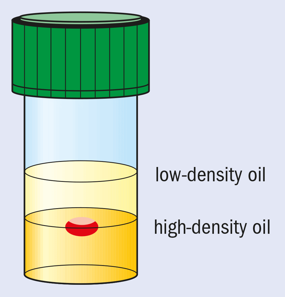
Another approach is to employ microgravity conditions to eliminate convection and sedimentation. This allows solely diffusion-driven conditions to be established. These are thought to be ideal for high-quality crystal growth and, in particular, the growth of crystals with weak intermolecular forces and lattice interactions. Microgravity conditions for crystallization can be partially mimicked by growing crystals in oil.
This method, pioneered at Imperial College, involves “containerless” growth with the drop containing the protein and the crystallizing agents suspended between two different oils with carefully chosen densities (see figure). The bottom layer is a high-density (1.27 g cm–3) fluorinated silicone fluid and the top layer contains low-density standard silicone fluid (0.92 g cm–3).
The actual X-ray data collection, if performed at or near room temperature, can lead to serious radiation damage to the crystals due to their high water content. The X-rays generate free radicals, which travel through the solvent channels that criss-cross the crystal and attack the intermolecular contacts between the adjacent protein molecules. This eventually leads to the break up of the crystal. However, the channels can also be used to diffuse small molecules to the active sites on the protein. This forms the basis of the growing field of time-resolved macromolecular crystallography.
High-speed data collection and short wavelengths do reduce damage significantly but the best approach is to freeze the crystal by plunging it into liquid nitrogen or propane. It might be thought that the expansion of the water when it freezes into ice would damage the crystal, but this can be avoided if the temperature is reduced quickly enough. Elspeth Garman of the molecular biophysics department at Oxford University in the UK is a pioneer of these techniques, while Mike Glazer of the physics department at Oxford has developed liquid-nitrogen apparatus that is economic to run and that is now marketed by Oxford Cryosystems.
So far the best results have been obtained with very small crystals (less than 300 microns). However, this is not a problem because the high brilliance of synchrotron X-ray beams still allows excellent data to be collected from such small crystals.
Freezing changes the protein structure slightly and it is obviously important to take this into account when using such structures in the design of drugs that will have to work at room temperature. However, this slight complication is greatly outweighed by the reduction in radiation damage to the crystal sample.
Position-sensitive X-ray detectors are another area of active research. In the UK, for example, Oxford Instruments is leading the multi-million pound IMPACT (Innovative Microelectronic Pixellated Sensors and Advanced CCD Technology) programme to develop novel X-ray detectors that combine charge coupled devices and pixellated silicon detectors. The aim of IMPACT, which involves some of the UK’s most successful instrumentation companies working with the academic research community, is to develop new detectors for both commercial and scientific applications. Other efforts are centred at the CHESS synchrotron at Cornell University in the US, the University of California at San Diego and the Advanced Light Source at the Lawrence Berkeley National Laboratory, also in California.
The combination of a frozen protein crystal that is radiation hard, an extremely intense X-ray beam and a sensitive area detector such as a charge-coupled device (CCD) has opened up a new territory in determining protein structures. In Manchester, for example, we have worked closely with researchers at the CHESS synchrotron, along with structural biologists from the Weizmann Institute in Israel, to determine the structure of concanavalin A, a 25 000 molecular weight protein isolated from plants, at a resolution of 0.94 Å. Much research and development in this area has been conducted at the EMBL Outstation in Hamburg by Keith Wilson and Zbyszek Dauter.
These experiments also allow the structure and bonding details of many of the hydrogen atoms in the protein to be seen directly, even those bound in water molecules, traditionally a speciality of neutron protein crystallography. However, if we want to study the exchange of these hydrogen atoms with an aqueous medium – which is relevant in catalysis, for example – we need to use neutrons. At one time it was thought that proteins like concanavalin A were just too large to be studied with neutron protein crystallography. However, a pilot experiment at the Institut Laue-Langevin neutron source in Grenoble has shown that it is possible to collect enough data to study the proton exchange process if a range of neutron wavelengths and a large-area image plate detector are used. The experiment also demonstrates the wide range of techniques that are sometimes needed to understand the function of just one protein.
Proteins are also of considerable industrial and medical interest. For example, concanavalin A binds glucose, and the structure of this complex, also determined in Manchester, now forms the basis of a glucose-based biosensor for diabetics that is being developed by Hugh Jones of the University of Wales Swansea and industry. Similar plant proteins, known as lectins, might be able to prevent HIV infection by competing with the HIV virus as it tries to attach itself to cells. This approach is being investigated by Pierre Rizkallah of Daresbury and Colin Reynolds of Liverpool John Moores University in the UK.
There has been remarkable progress in protein crystallography in recent years. Indeed the determination of all of the protein structures in the human genome is now a realistic prospect. How long might this take? If we assume no further breakthroughs in techniques, it takes one or two days of beam-time at a synchrotron radiation source to acquire the diffraction data that will allow a typical protein structure to be determined. Twenty instruments working world-wide in a coordinated way would then require about 10,000 days or almost 30 years of work.
However, improved position-sensitive X-ray detectors could speed up this process enormously. For example, the pixel detectors would yield data sets for each of two wavelengths or crystals in matters of minutes, rather than hours or days.
Therefore, the determination of genome level structure determination might well take considerably less than 30 years, assuming that we can grow the crystals fast enough. It would have vastly important consequences for improved pharmaceutical design and the understanding of genetic diseases. Overall this would be physics, with chemistry and biophysics, applied at its best.