Ferroelectric materials have a permanent electric dipole, an internal separation of the centres of positive and negative ionic lattices, that can be flipped by applying an electric field. They also undergo a structural change at a material dependent temperature. known as the Curie temperature, above which this dipole behaviour disappears. Despite having permanent dipoles, ferroelectrics are insulating materials. These properties make them valuable in technologies such as sensors, actuators, and memory devices.
In this work, the researchers study the band gaps of ferroelectric materials to better enable their use in energy conversion, catalysis, and optoelectronic devices, where understanding light absorption and electron behaviour is essential. Traditionally, the band gap in ferroelectrics has been treated as a single number. However, ferroelectrics are not conventional semiconductors. They contain localized charges, polarons, internal dipoles, and structural disorder. These features give rise to three distinct band gaps, not one.
There is the intrinsic fundamental band gap, defined as the ground state difference between the fully occupied valence band and the completely empty conduction band. The smaller optical gap is associated with light induced transitions involving bound electron-hole pairs (excitons), and the even smaller transport gap associated with electrical conduction via localised electronic carriers.
In this study, the authors determine the fundamental, optical, and transport gaps using X‑ray photoelectron spectroscopy, optical spectroscopy, and electrical conductivity measurements, respectively, for NBT‑6BT and NaNbO₃. The fundamental gap values are further supported by DFT calculations. Because these three gaps differ by about 1 eV or more, different experiments have actually been probing different gaps all along, meaning past optical and electrical results were often compared incorrectly, leading to widespread misinterpretation. The conclusion establishes that ferroelectrics possess three fundamentally different energy gaps, explains why they differ, provides a framework for measuring them, confirms their values theoretically, and highlights why this distinction is crucial for designing future energy and electronic technologies.
Tests of fundamental physics that were previously impossible could become a reality thanks to a new way of producing extremely intense beams of light. Using a state-of-the-art high-power laser, researchers at the University of Oxford, UK demonstrated that they could dramatically increase the efficiency of a nonlinear optical technique called relativistic harmonic generation. According to the team, this increase could herald a paradigm shift, making it possible to achieve hitherto unheard-of electromagnetic field intensities in the laboratory.
The theory of quantum electrodynamics (QED) predicts that at very high intensities, light can interact with the vacuum, converting light energy directly into matter. “If we can achieve such intensities, we could test theories about the fundamental nature of the universe,” says Robin Timmis, who led the new study. “However, doing so requires a laser system a million times more intense than those currently available.”
Relativistic harmonic generation
In the new work, Timmis and her colleagues in Peter Norreys’ group at Oxford used the Gemini laser at the UK Science and Technology Facilities Council’s Central Laser Facility (CLF) to generate coherent extreme ultraviolet (XUV) and X-ray photons via relativistic harmonic generation. They began by firing high-frequency, ultrashort, sub-picosecond (10-12 s) laser pulses onto a solid glass target. This creates a plasma that acts like an oscillating mirror, and Timmis likens the next step to shining a flashlight at this mirror while it is rushing towards you at near-light speed – a concept known as “Einstein’s flying mirror”. The result is that the light reflected from the plasma becomes compressed, and gains intensity.
Working with researchers from Brendan Dromey‘s group at Queen’s University Belfast in Northern Ireland, the team used a process called coherent harmonic focus to concentrate this light into a region as small as a few nanometres across. This step may have boosted the light beam’s intensities as high as 1023 W cm−2, although Timmis acknowledges that this is an estimate based on previous theoretical simulations, as the team was unable to measure it directly.
“If confirmed with further experiments at Gemini, or indeed even larger facilities, we may have made the most intense source of coherent light ever,” says Timmis, who received this year’s Institute of Physics Culham Thesis Prize in part thanks to this work, which is described in Nature. “The energy in our XUV beam was over three orders of magnitude brighter than previous measurements,” she adds. “By resolving a long-standing gap between theoretical expectations and experimental results, we confirmed the required energies to support a coherent harmonic focus and therefore offer a substantial boost in intensity above that of the original laser pulse.”
Towards the next generation of extreme electromagnetic field studies
According to the researchers, these results demonstrate that there is a realistic experimental pathway to next-generation laboratory studies of extreme electromagnetic fields. In particular, they say that the quantum critical field for QED tests, which is known as the Schwinger limit and has a value of >1016 V cm−1 or >1029 W cm−2, is now open, paving the way towards all-optical studies of the quantum vacuum. As well as fundamental physics, Timmis says that more efficient harmonic generation could also have applications in ultrafast imaging of physical and biological systems, photolithography and fusion science.
The Oxford team is now analysing data from a follow-on experiment at the CLF that will guide their next steps. “We will be shortly publishing results about a new harmonic beam that we have discovered on that run,” reveals Timmis, “and future studies will focus on actively controlling the coherent harmonic focus and directly measuring its intensity.”
Researchers at Stanford University in the US have found a way to generate light deep within living tissues, potentially leading to new forms of gene and cancer therapies. The proof-of-concept approach uses ultrasound to trigger luminescence in nanoscale particles travelling through the bloodstream, and it has already been tested in tissue-mimicking “phantoms” and live mice. However, its developers caution that human trials are still some way off.
Light has numerous applications in medicine and biological research. It is widely used, for example, to stimulate cell growth and in photodynamic therapies for skin and eye conditions, as well as certain types of cancer.
The problem is that many potentially useful wavelengths of light are easily scattered by tissues and become attenuated over relatively short distances. This means they cannot penetrate very far into the body without help from invasive methods such as removing overlying tissue or inserting/injecting optical implants and light-emitting nanoparticles into the target area.
Sound and light
The new work by Stanford materials scientist and engineer Guosong Hong and colleagues involves nanoparticles made from a ceramic material with the chemical formula Sr4Al14O25:Eu,Dy. This material is mechanoluminescent, meaning that it emits light when subjected to mechanical stresses and deformations. In Sr4Al14O25:Eu,Dy, these mechanoluminescent effects can be induced by exposing the material to sound waves, which penetrate more deeply into tissue than light waves.
The Stanford researchers began by coating their nanoparticles with a biocompatible film. They then suspended the particles in a solution and injected the resulting colloid into the veins of mice. Thanks to the rodents’ vascular systems, the particles soon travelled to all parts of their bodies.
The researchers then showed they could make the nanoparticles emit blue light with a wavelength of 490 nm simultaneously in multiple locations (such as the brain, gut, hindlimb and spine) by applying sound waves to different parts of the mouse’s body. In addition, they showed they could create precise patterns of in-situ light generation throughout the three-dimensional volume of the animal, controlled over distances of 100 to 200-μm in the focal region. The ultrasound can also be used as a scanner to define where the light is generated.
A host of applications
The team picked the 490 nm wavelength because it has many applications, including neuron modulation and photodynamic cancer therapy. However, applying the same technique to different materials could produce other useful wavelengths, too. Indeed, Hong and his colleagues are exploring the possibility of using materials that emit ultraviolet light, which has antiviral and antibacterial properties.
The researchers say their approach is broadly applicable to virtually all therapeutic modalities that requires light to be delivered deep within the body, including optogenetics, phototherapy and photo-switchable gene editing. This last technique currently suffers from off-target effects, but the researchers say that by pairing light-producing nanoparticles with a light-activated gene-editing system, they may be able to use ultrasound to turn gene editing on and off in localized areas of the body.
“The overarching theme of my lab’s research is to develop new strategies to deliver and receive light throughout the body in its native, living state,” Hong tells Physics World. “In 2024, we reported on a method to render living tissue transparent using strongly absorbing dye molecules. In the present study we have taken a complementary approach: rather than modifying how light propagates through tissue, we leverage the intrinsic penetrative capability of ultrasound, together with the pervasive reach of the circulatory system, to generate light directly within deep regions of the body.”
Reporting their work in Nature Materials, the researchers are now working to integrate their approach with other light-activatable control systems, including photo-switchable Cas9 gene editing in collaboration with Michael Lin’s lab at Stanford. In parallel, they hope to develop alternative mechanoluminescent materials that will break down safely in the body. While the materials studied in this work did not seem to show adverse effects in mice, they also did not break down quickly, and the researchers say they could accumulate in organs such as the liver.
“What we’re demonstrating here is a proof-of-concept showing that you can produce light emission in a programmable manner deep within the body,” Hong says. “If we can replace the material with one that is safer to be used in humans, that will start to pave the way for clinical applications.”
Quantum technologies are undoubtedly going to have a large impact on our world, potentially revolutionizing everything from healthcare and the environment, boosting the economy and helping with large-scale optimization challenges. But for them to deliver on these many promises, it will be vital for many countries to train and build a quantum-ready workforce.
There are four pillars to the quantum sector – quantum computing; quantum simulation; quantum communication; and quantum sensing and metrology. But in each case there is a lack of trained individuals who can take on jobs across the board. Indeed, statistics in both the UK and the US suggest there is only one qualified worker for every three quantum jobs. With governments continuing to invest lots of money into national quantum programmes; a growing number of new quantum start-ups being launched; and ever more multi-national firms zoning in on quantum, the shortage of those with the right skills to work across the sector is expanding.
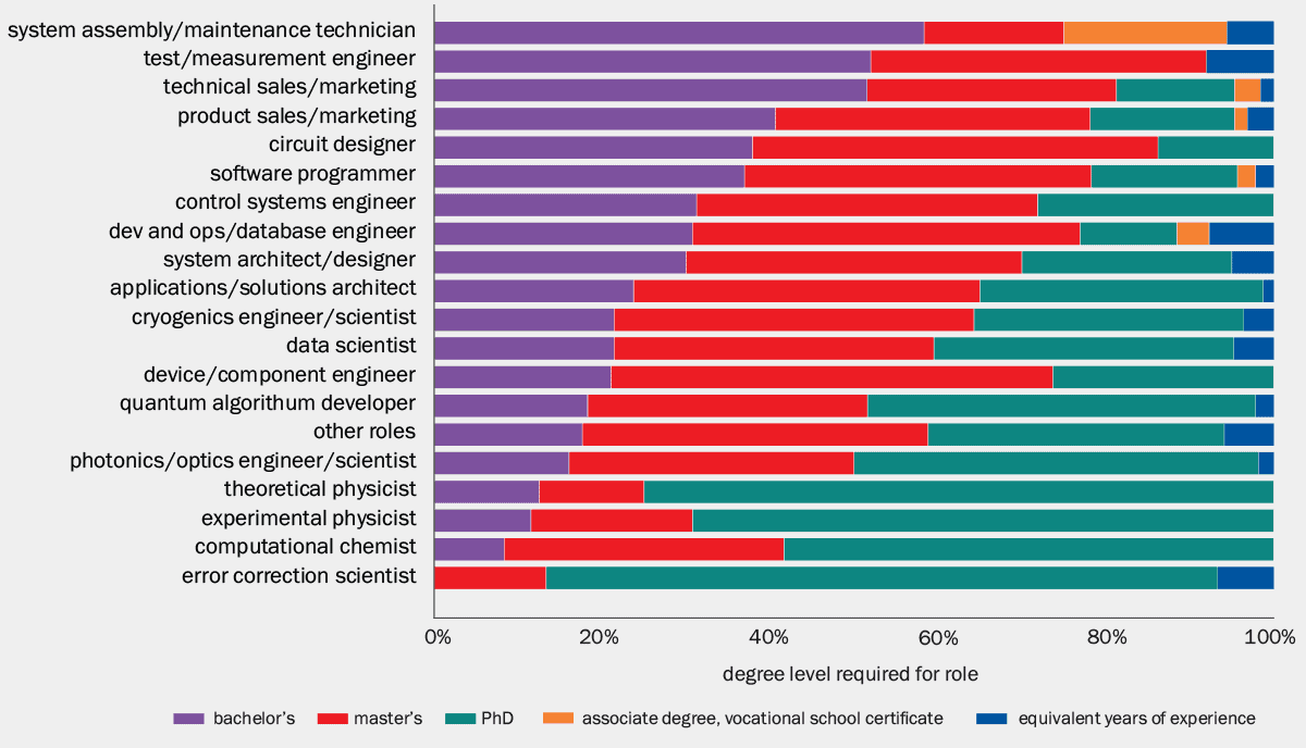
The Colorado School of Mines in the US is now trying to remedy this situation by launching the country’s first bachelor-level quantum systems engineering degree programme, due to start this autumn. An undergraduate degree specializing in quantum and systems engineering might, at first glance, seem odd. But 2021–2023 data from the Chicago Quantum Exchange show that 55% of quantum tech jobs only require a BSc or two-year associate degree. For instance, roles that ask for just a BSc include systems assembly and maintenance, measurement engineers, technical sales and marketing.
“Industry demand especially values engineers with a systems-level understanding of quantum devices, and there is also a need for quantum technicians who can build and maintain quantum hardware,” says Frédéric Sarazin, director of the quantum programme at Colorado School of Mines. As the first standalone bachelor’s degree in quantum systems engineering in the US, the programme is designed specifically to supply industry-ready graduates.
True requirements Distribution of degrees needed for different job roles in the quantum industry. (CC BY 4.0 IEEE Transactions on Education65 592)
The main focus for Sarazin and colleagues was to bring into the programme key aspects of systems engineering – which involves understanding and overseeing all aspects of a complex system, from its inception through to practical production, and even managing the final product. The goal: to help companies get their products and technologies out of the lab and into the marketplace. Rather than focusing on isolated components, systems engineers are trained to understand how complex technologies behave as integrated entities.
“A quantum computer, for example, is more than just its qubits,” says Sarazin. “It’s cryogenics, optics, electronics, control software, signal processing and the user interface, all interacting with each other.” Companies are keen to hire people who can understand and help develop their quantum product as an end-to-end system, bridging the gap between the physics and engineering aspects, as well as making sure the end product is robust, scalable and manufacturable.
The physics may be what Sarazin calls the “secret sauce” – but turning it into a device that is reliable, manufacturable and maintainable is an engineering problem “with a quantum flavour to it”. “What companies want is people who understand the product as a system, from beginning to end,” Sarazin explains.
Quantum hotspot
Colorado, in America’s mid-west, is a quantum innovation hotspot, with quantum companies employing more than 3000 people across the state. To develop the new programme, Sarazin and colleagues carried out an extensive consultation process with companies, institutions and organizations that all look to hire quantum engineers, to get a clear idea of the skills that students should have at the end of their course. They also collaborated with Elevate Quantum – a consortium of 120 organizations advancing quantum workforce development and commercialization in Colorado, New Mexico and Wyoming – to design an interdisciplinary course that will integrate physics, electrical and mechanical engineering, computer science and engineering design.
While the students will learn plenty of foundational quantum physics, they won’t cover the full curriculum of a traditional physics degree. “You’d be talking about a six-year degree if we covered everything,” says Sarazin. Certain advanced topics, such as quantum error correction, remain overwhelmingly in the domain of PhD-level jobs and so are deliberately excluded.
The lab is meant to be a signature experience. It’s where students start interacting with industry in a meaningful way
A key feature of this degree will be hands-on practical engineering experience in the lab. Plans are under way to build a dedicated quantum device laboratory for the students, allowing companies to bring in their tech and partner with the on-campus facilities. “The lab is meant to be a signature experience,” says Sarazin. “It’s where students start interacting with industry in a meaningful way.”
That connection is reinforced through internships and a year-long design project in the final year, with project topics supplied directly by quantum companies. “The junior-to-senior year is when internships really matter,” explains Sarazin. “That’s often what leads directly to a job.”
Future prospects
Although the programme is firmly industry-focused and aims to get graduates straight into the job market, students can progress to the Colorado School of Mines’ existing master’s programme in quantum engineering, launched in 2020. “At the bachelor’s level, you’re building breadth,” says Sarazin. “If students want to specialize further, they absolutely can.”
Many of the skills that the students will develop – from electronics and embedded systems to control software and algorithms – are highly transferable too. “Looking beyond the quantum sector, our systems engineering students will have acquired a set of skills that is highly applicable in other industries,” says Sarazin.
The first cohort will likely be around 15–20 students this year. Looking ahead, Sarazin has a clear benchmark for success: “a near-100% placement in industry at the end of the degree – that’s what we’re aiming for”.
Beyond that, success will mean continuously refining the programme in response to industry feedback. “This isn’t static,” Sarazin says. “If companies tell us something needs adjusting, we want to respond.” For students still hesitant to take the leap into a specialized BSc or the quantum sector, Sarazin’s message is clear: quantum careers are here to stay and the direct path into the industry is starting earlier than ever before.
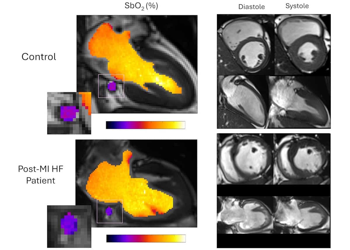
MRI measurements Representative blood oxygen saturation (SbO2) maps and left ventricle (LV) geometry images from a control participant and a patient with heart failure, demonstrating reduced coronary sinus SbO2 and impaired LV contractility in the patient. The colour scale indicates SbO2 of 20 to 100%. (Courtesy: Ting Huang et al. Sci. Trans. Med. 18 eady6269 (2026))
The amount of oxygen that a heart consumes is a key indicator of its health. If the heart is not receiving or using enough oxygen, heart tissue can be damaged, contributing to future heart failure.
With abnormal myocardial oxygen consumption an indicator of potential cardiac dysfunction, its measurement could help in the early detection and treatment of heart failure. And as one in four individuals are likely to develop heart failure in their lifetime, this is of critical importance. But measurement of myocardial oxygen consumption is not a simple process. The gold standard for determining the heart’s oxygen use is cardiac catheterization. But this test – which involves threading a catheter from a patient’s neck or groin into the coronary sinus (CS), the largest coronary vein – is highly invasive, time-consuming and comes with a level of risk.
A new MRI technique may soon offer a rapid, non-invasive alternative. Developed by an international research team headed up at Cedars-Sinai Health Sciences University, the high-resolution MRI method can assess the heart’s oxygen consumption in just three minutes. In an initial study of 22 patients with heart failure, reported in Science Translational Medicine, the team validated its accuracy, feasibility, performance and repeatability.
Principal investigator Hsin-Jung Yang is director of cardiac imaging research in the Biomedical Imaging Research Institute at Cedars-Sinai Medical Center.
MRI is sensitive to blood oxygenation via the blood oxygen level–dependent (BOLD) signal, originally developed for mapping brain activity. Use in the heart remains challenging, however, due to the need for complex calibration, motion sensitivity and long acquisition times. Hsin-Jung Yang, of the Biomedical Imaging Research Institute at Cedars-Sinai, and collaborators overcame these obstacles by developing a rapid, self-calibrated cardiac MRI framework that enables free-breathing blood oximetry (measurement of blood oxygen saturation) in the CS and quantification of whole-heart myocardial oxygen extraction, without requiring contrast agents or pharmaceutical stress.
The researchers’ primary objective was to determine the accuracy and precision of MRI-derived measurements of CS blood oxygenation, compared with those obtained by invasive CS catheterization. They also aimed to perform non-invasive quantification of global myocardial oxygen consumption and myocardial oxygen efficiency, with comparisons between healthy controls and patients with heart failure.
To achieve this, they developed a motion-resolved reconstruction algorithm for cardiac BOLD MRI that enables clear imaging of the moving heart during breathing and heartbeats. The team first validated the method in pigs, and then applied it to a group of 22 patients with heart failure and a history of previous heart attack, as well as 11 healthy volunteers.
The researchers acquired clinical cine images to define the cardiac anatomy, localize the CS and measure ventricular function for estimating the oxygen–mechanical work coupling efficiency. Using this approach, they identified impaired myocardial oxygen consumption in the patient group, including those with preserved ejection fraction (how much blood the left ventricle pumps out with each contraction, a low value of which can indicate a heart problem). The finding that impaired oxygen consumption was measurable even before detectable structural or functional decline may facilitate the early detection of cardiac dysfunction.
The researchers note that their self-calibrated MRI framework directly addresses the difficulty of performing quantitative oximetry of the CS – a mobile blood vessel that undergoes marked displacement throughout the cardiac cycle. “Our framework directly addresses these challenges with a continuous, free-breathing, motion-resolved 3D acquisition that retrospectively sorts data across cardiac and respiratory phases, ensuring stable CS tracking despite its complex motion and size variation,” they write.
By eliminating the dependence on gating and calibration, the method could be applied across diverse clinical populations, including those with arrhythmias, intolerance of breath-holding or physiologic stress, for whom conventional gated acquisitions are unreliable. The team suggests that the framework also holds promise for extending oxygen consumption imaging to other moving organs, such as the liver and kidney, and that in the future, the motion-resolved BOLD framework could be applied to tissue-based quantification.
The researchers are performing ongoing clinical studies to evaluate the MRI technique in aortic stenosis (narrowing of the aortic valve) and hypertrophic cardiomyopathy (thickening of the heart muscle), where altered oxygen extraction and metabolic efficiency have revealed disease severity, risk and treatment response beyond conventional imaging.
More broadly, the Yang Lab is extending this approach to characterize oxygen utilization in all cardiometabolic diseases and associated emergent therapies, with the goal of noninvasively defining myocardial energetic supply–demand balance, identifying therapy–response phenotypes, and monitoring disease progression and metabolic remodelling over time.
“By enabling a fast, non-contrast, non-ionizing radioactive method for measuring cardiac oxygen metabolism, [this MRI method] can unlock frontiers for early diagnosis, personalized therapy, and the development of next-generation cardiometabolic treatments to combat the global heart failure epidemic,” the team concludes.
An event was held yesterday – attended by senior officials including CERN director-general Mark Thomson and Dario Gil, undersecretary for science at the US Department of Energy (DOE) – to commemorate the start of moving 4.5 million kilograms of steel beams underground that will be used to hold DUNE’s detectors in place.
In February 2024, excavation work finished on two huge underground spaces for DUNE. Located 1.6 km underground at the Sanford Underground Research Facility in South Dakota and are some 150 m long and seven storeys tall, the spaces will be used to house DUNE’s four neutrino detector tanks that are each filled with 17,000 tonnes of liquid argon and cooled to 88 K.
When complete in 2031, DUNE-LBNF will study the properties of neutrinos in unprecedented detail, as well as the differences in behaviour between neutrinos and antineutrinos.
DUNE will measure the neutrinos that are generated by Fermilab’s accelerator complex, which lies around 1300 km away just outside Chicago.
The cryostat materials, which have been contributed by the CERN, are now scheduled to be moved underground and installed in the next few months.
“Today represents the start of a pivotal phase for DUNE, the development of the far detector structures in South Dakota,” noted Fermilab director Norbert Holtkamp. “Our focus remains on safety, quality and schedule — in that order — to ensure we successfully deliver on behalf of the US Department of Energy, our nation and the world.”
Physicists have determined the mass of the W boson with the highest precision yet by analysing more than a billion proton collision events at CERN’s Large Hadron Collider (LHC). The new result confirms a prediction from the Standard Model of particle physics while refuting a comparably precise measurement made by Fermilab’s CDF Collaboration in 2022. This is significant because the older measurement, which used data from the defunct Tevatron collider, differed from the Standard Model’s predictions by seven standard deviations, suggesting that the W boson might be far heavier than the model allows.
The W boson is one of two elementary particles that acts as a carrier for the weak force (the other is the Z boson). As one of the four fundamental forces in nature, the weak force is what allows protons to change into neutrons (and vice versa), making it the driving factor behind radioactive decay and nuclear fusion. Precise measurements of the W and Z boson basses are therefore important for understanding these processes as well as for testing the Standard Model.
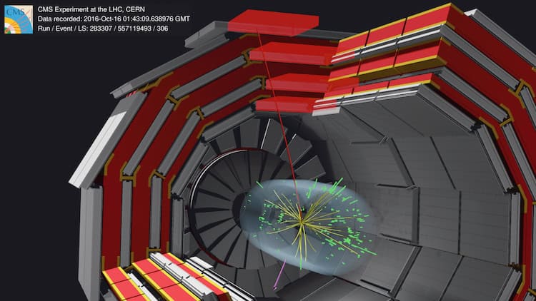
Particles incoming: An event picture showing how the researchers present their recordings digitally to illustrate the detector readings. (Courtesy: C Paus)
While physicists have measured the mass of the Z boson to an extremely high precision of 22 ppm (or 2.0 MeV), measuring the mass of the W boson with the same exactitude has proven more difficult. The main hurdle is that the W boson cannot easily be detected in colliders such as the LHC because it decays almost instantly. Scientists can look for its decay products instead, but that, too, is awkward. In one important channel, for example, it decays into a neutrino and a muon – and neutrinos are even more elusive than W bosons.
A fading mystery
In the new work, CERN’s Compact Muon Solenoid (CMS) Collaboration studied more than a billion proton collision events produced at the LHC in 2016. Amongst these, they identified 100 million as producing a W boson that decayed into a neutrino and a muon.
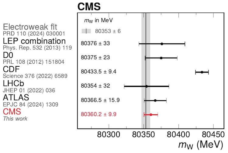
A comparison of the most recent W boson mass result split by experiments (LEP is four experiments combined). Courtesy: C Paus
By analysing these events and simulating all the possible scenarios that could produce them, they measured the mass of the W boson to be 80360.2 ± 9.9 MeV. This is significantly less than the CDF Collaboration’s measurement, but it agrees with other previous experiments. Importantly, it also lies within the range the Standard Model predicts, leaving the CDF result – the most precise measurement before this one – looking like an outlier.
“If you take the CDF measurement at face value, you would say there must be new physics beyond the Standard Model,” says Christoph Paus, a physicist at the Massachusetts Institute of Technology (MIT) in the US and one of the lead investigators of the CMS Collaboration. “And of course, that was the big mystery.”
Now that the new, even more precise measurement agrees well with predicted values for the W boson mass, that mystery is fading, Paus tells Physics World.
Some physicists may find this disappointing. However, study lead author Kenneth Long, who was a senior postdoc in MIT’s Laboratory for Nuclear Science at the time and has since moved to a research position in Lyon, France, says the new result is “just a huge relief to be honest” and “a strong confirmation that we can trust the Standard Model”.
A starting point for precision measurements
To obtain their result, the CMS researchers needed to measure the momentum of the muon and use it to infer the W boson’s mass. This is possible for two reasons. The first is that in the W’s rest frame, its decay energy is shared roughly equally between its two daughter particles (the muon and the neutrino). The second is that muons are charged leptons, and the strong magnetic field inside the CMS detector makes them travel in a path whose curvature is a function of their momentum.
“The momentum is different to the mass, of course, but is strongly correlated with it,” explains Paus. “The challenge is therefore to track the path of the muon and every possible interaction it could have with other particles and its surroundings to estimate a value for its initial momentum.”
The CMS experiment had long planned on doing such a measurement, but it took a while to set up. Now that the measurement is complete, Paus, whose MIT group joined the W boson mass analysis effort in earnest at the end of 2020, describes it as an important starting point for the collaboration. He explains that the result proves it’s possible to measure the W boson in what he calls a “high pile-up environment”, meaning one where many proton-proton collisions overlap in a single recording, without using the Z boson mass as a calibration (as was previously done in analyses at hadron colliders). “It has put the CMS experiment finally on the map for an electroweak precision measurement of this kind,” he says.
The CMS researchers are now collaborating with experimentalist colleagues at CERN’s ATLAS and LHCb detectors, as well as their theorist partners, in hopes of setting a new standard in electroweak precision physics. Their measurement is published in Nature.
Nuclear fusion powers the Sun, and scientists and engineers have long been trying to harness the process to generate clean energy. While much progress has been made, the commercially-viable generation of fusion energy remains elusive.
One important challenge is developing a range of specialized materials that can contain an extremely hot, radiation-emitting plasma in close proximity to ultracold superconducting magnets.
Our guest this week is Jacob John of the UK Atomic Energy Authority, who studies how radiation damages materials. In conversation with Physics World’s Matin Durrani, he talks about the near-oxymoronic materials requirements for fusion reactors and how they can be met.