Image data offer functional clues for lung radiotherapy

Lung radiotherapy treatment plans

Precision is everything in radiotherapy treatments, with the overriding objective to deliver the radiation dose directly to the tumour without causing any damage to healthy tissue and the surrounding organs. When treating the lung, for example, exposing the parts that are responsible for gas exchange to ionizing radiation can trigger a range of respiratory issues, ranging from short-term inflammation and shortness of breath to chronic conditions that can cause permanent scarring of the lung tissue.

“The lung is an important organ-at-risk, and radiation-induced toxicity is our main concern when treating patients with lung tumours,” says Florian Putz, a senior radiation oncologist at the University Hospital Erlangen in Germany. “The standard clinical practice is to minimize the dose across the entire organ, but that doesn’t take account of any spatial variation in the ventilation provided by different parts of the lung.”

Putz points out that the most functional parts of the lung – the areas that efficiently extract oxygen from the air we breathe – are not always equally distributed. Localized tissue scarring can compromise the gas-exchange process in some places, while any inflammation can constrict the airways in particular areas of the lung. Taking account of these spatial variations would enable clinicians to spare the parts of the lung that contribute most to a patient’s breathing capacity, and offers the potential to deliver more dose to areas where ventilation is already suppressed.

Putz has been investigating whether such spatial information on pulmonary function could be retrieved from the data recorded in time-resolved CT scans. Such 4DCT scans are routinely acquired during thoracic treatments, primarily to record the movement of the tumour as the patient breathes in and out, and a novel tool built into the syngo.via RT Image Suite from Siemens Healthineers uses this data to calculate and image the ventilation in the five distinct lobes of the lung. “This lung ventilation tool would enable us to spare the lobes that contribute most to the patient’s breathing capacity, and offers the potential of escalating the dose that is delivered to the lobes that are not so functionally relevant,” explains Putz.

Inside the tool is an automated deep-learning technique that uses the 4DCT images to segment the five lobes of the lung – first when the patient has taken a full inhalation and then when all the air has been expelled. The ventilation in each lobe is then calculated from the difference in air volume between those two states, and then expressed as percentage value by normalizing with the air volume at maximum exhalation.

“The segmentation gives us the total volume of the lung in each state, and then we calculate the air volume from a simple model that treats the lung as a linear combination of air and water,” explains Christian Möhler, product manager for radiotherapy imaging software at Siemens Healthineers. “This straightforward mathematical approach is robust to uncertainties, and we can use the percentage values to provide a colour-coded view of the ventilation in each of the five lobes.”

Other methods have been proposed to extract ventilation information from time-resolved CT, in many cases providing a granular view down to the level of individual voxels. In this case, however, each data point in the CT image must be correlated between the inspiration and expiration states, which can introduce inaccuracies when trying to map the intricate airways and blood vessels within the lung. In contrast, the algorithm developed by Siemens Healthineers avoids the need for this complex registration process, instead relying only on the segmentation of the five lobes that are produced for each breathing state.

“We need to avoid any errors that could lead to wrong treatment decisions, which means that the algorithm needs to be robust,” comments Putz. “The lobes also tend to be quite homogeneous, which means that the ventilation across the spatial compartment of each one tends to be quite similar.”

Putz has been working with medical physicist Juliane Szkitsak to compare the information extracted from the CT lung ventilation tool with the data from standard breathing tests that exploit spirometry to assess the overall pulmonary function of both lungs. From the ventilation metric provided by the software tool he calculated the volume of air across all five lobes of the lung, allowing a direct comparison with the results from these spirometry tests. “There was a high correlation between the two, which suggests that the tool is working as it should,” he says.

In a further evaluation at University Hospital Erlangen, Szkitsak replanned the treatment of six patients using information from the CT lung ventilation tool. “The new treatment plans avoid the healthy parts of the lung and only treat through the lobes that the ventilation data show might already be impaired,” she explains. “The additional information from the ventilation tool made the optimization more complex, but I think it would become quicker and easier as you get more familiar with it.”

However, Szkitsak also points out that for this small selection of patients it was difficult to assess the benefits of using the tool, since the complexity of thoracic treatment plans and the need to avoid other organs-at-risk makes it difficult to produce comparable plans. She also found that in some cases the new treatment plans resulted in the radiation beam passing through more of the lung, which was a cause for concern among the clinical team at Erlangen. “Usually we avoid as much lung volume as possible by, for example, minimizing the distance between the skin and the tumour,” she explains. “With the plans produced using the ventilation algorithm we sometimes had to go through the lung in a different way, with the result that more of the lung was exposed to radiation.”

With no clear evidence to show that the ventilation data can reduce the risk of damaging the lung, the clinicians at Erlangen have concluded that it is too early to change their treatment strategy. “While there is a growing consensus in the literature that CT lung ventilation produces valid results, all the physicians at our hospital need to be convinced that it can provide a benefit for patients without putting any of them at risk,” says Putz. “Additional studies and larger evaluations will be needed, while widespread acceptance will most likely require clinical trials that show reduced pulmonary toxicity in treatments that exploit such a tool.”

While the ventilation information might not yet influence the initial treatment planning process, Putz believes that it could help clinicians to monitor lung function during multiple rounds of treatment. Such regular monitoring could augment the standard breathing tests that are usually performed before treatment starts, helping clinicians to detect early signs of toxicity and even to adapt their treatment plans to take account of any changes in ventilation.

“We are moving towards adaptive radiotherapy, in which we take multiple scans as the treatment progresses and then update the treatment plan to reflect any changes in the body,” explains Putz. “Using this tool we have seen changes in the spatial distribution of the ventilation during treatment, for example in patients where pulmonary effusion caused fluid to accumulate in the lung, and having this information would enable us to update our treatment plan and deliver better outcomes for the patient.”
My Favourite Qubit: quantum simulation and computation with superconducting qubits

Want to learn more on this subject?

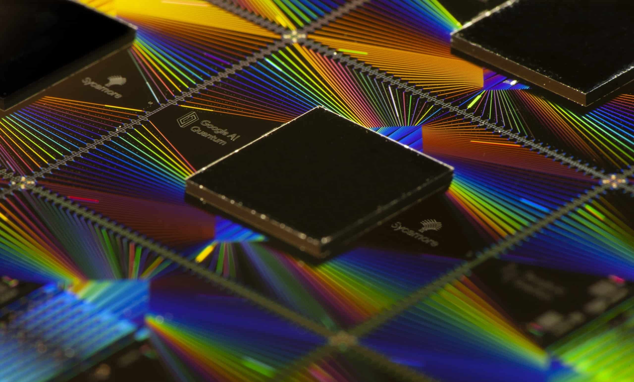
In recent years superconducting qubits have become one of the leading platforms for quantum computation and simulation. In 2019, the Google team demonstrated that a quantum processor could perform certain computational tasks exponentially faster than a classical computer. Going beyond this milestone, we utilize these Noisy Intermediate Scale Quantum (NISQ) processors to study nonequilibrium quantum dynamics and simulate quantum phases of matter. After introducing these processors, I will present some of our recent works.

Want to learn more on this subject?

Pedram Roushan received his BS in physics and mathematics from University of Pittsburgh in 2005 and his PhD in physics from Princeton University in 2011. During his PhD, he studied metal-insulator phase transitions and performed the first scanning tunneling microscopy of topological insulators. After postdoctoral studies at UC Santa Barbara, he joined Google quantum AI. The current focus of his research is on simulating novel condensed matter and non-equilibrium physics with superconducting quantum processors.


This webinar is the first in our ‘My Favourite Qubit’ series. Why not sign up for our other webinar? Even if you’re not able to join the live event, registering now enables you to access the recording as soon as it’s available.


My Favourite Qubit: the atomic ion

Want to learn more on this subject?

Internal energy levels of a single atom represent nature’s perfect qubit – the quantum two-level system that is the building block of quantum computing technology.

At the Duke Quantum Center, we trap single atomic ions using electric fields and manipulate the qubits using lasers to perform quantum gates.

In this webinar, Crystal Noel will describe how we start with single atoms and build a fully programmable quantum computer.

Want to learn more on this subject?

Crystal Noel is an assistant professor in electrical and computer engineering and physics at Duke University, where she is part of the Duke Quantum Center. Crystal received her BS from MIT in 2013, and then moved to UC Berkeley for her graduate studies with Prof. Hartmut Haeffner. After completing her PhD in trapped ion quantum computation in 2019, she worked with Christopher Monroe at University of Maryland as a postdoc. Crystal started at Duke in 2021 as a research scientist before joining the faculty in 2022. Her research spans the field of trapped ions from device engineering and photonics integration to quantum computing systems engineering to high-level algorithms and applications.

This webinar is the second in our ‘My Favourite Qubit’ series. Why not sign up for our other webinar? Even if you’re not able to join the live event, registering now enables you to access the recording as soon as it’s available.


Chiral detection of molecules gets a laser-driven boost

Physicists in the UK have developed a new and highly efficient way of detecting the chirality or “handedness” of molecules using tailored laser fields. The technique could improve the process of drug development in medicine and might also become the basis for compact devices that rapidly separate left- and right-handed molecules.

“Chirality is a universal type of asymmetry that arises naturally in many areas of science,” explains David Ayuso, a physicist at Imperial College London who led the research. “In general, an object is chiral when it is different from its mirror image – with our hands being the typical example.”

Just as a chiral glove would either fit a left hand or a right hand, but not both, the two superimposable, mirror-reflected versions of a chiral molecule (called enantiomers) behave very differently when they interact with another chiral entity such as circularly polarized light or a different chiral molecule. Since most biomolecules are chiral, and some of these different behaviours have detrimental or even toxic effects, reliable methods of detecting, quantifying and manipulating molecular chirality are highly desirable in the context of biochemistry and pharmaceuticals.

A fundamental limitation to overcome

Conventional optical methods for measuring chirality in matter rely on the helical – and thus chiral – pattern that the polarization of circularly polarized light “draws” as the light propagates through space. The problem is that the pitch of this helix is determined by the light’s wavelength, which is orders of magnitude longer than the molecules that interact with it. Consequently, the molecules “see” the helix as a simple planar circle – a non-chiral structure. As a result, the light-molecule interaction is only very weakly sensitive to the molecule’s handedness, with sensitivities usually below 0.1%.

Ayuso and colleagues have been working on several approaches over the last few years to overcome this fundamental limitation. “The key is to stop relying on the chiral helix that circularly polarized light draws in space, but instead try and create new optical fields in which the tip of the electric field vector (or the field’s ‘arrow’) of the laser creates a chiral structure in time,” says Ayuso. “In this way we create light that encodes chirality in time rather than space. The molecules therefore no longer perceive a circle but rather a chiral temporal structure.”

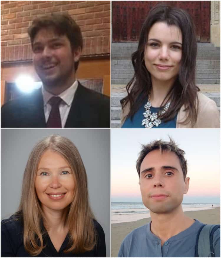
Photos of the four research team members

Light that encodes chirality in time is efficient at driving chiral electronic currents inside the molecules, he tells Physics World. These currents then interact with the molecules’ chiral structures (their natural “corkscrew”) in a way that is highly enantio-sensitive – that is, strongly influenced by the molecule’s handedness. “In this way, we can ‘force’ one of the two versions of a chiral molecule, for example, the left-handed one, to emit bright light at new optical frequencies, while the right-handed one remains dark,” he explains. “This allows us to detect the chirality of a molecule with 100% efficiency.”

Devices for efficient chiral recognition

According to the researchers, being able to drive strongly enantio-sensitive signals with lasers could aid the development of compact devices that recognize and manipulate chiral molecules in a highly efficient way. One example might be a pair of chiral optical tweezers that distinguishes between left-and right-handed molecules in space.

The Imperial College team is now collaborating with experimentalist colleagues Mary Matthews and Jon Marangos to develop the technique further. As part of this effort, a PhD student in the team, Rose Piccuito, is applying “the unique laser capabilities available at Imperial” to the task, Ayuso says. “In her experiments, she uses femtosecond laser pulses (that is, ultrashort pulses that last just around 10-15 seconds). The goal here is to image the chiral molecules not only with extremely high chiral sensitivity, but also at the natural timescales of electronic motion,” he explains.

Another interesting direction, he adds, would be to bring these ideas to the nanoworld. “We want to take advantage of the unique opportunities enabled by nanophotonic technology to shape light for highly enantio-sensitive imaging and manipulation of chiral molecules.”

The method is described in Science Advances.

Spinning a sustainable fashion revolution: meet the physicists turning wood into clothes

You might be somewhat shocked to know that, according to a recent European Union (EU) study, worldwide fashion and clothing is responsible for 10% of global CO emissions – that’s more than international flights and maritime shipping combined. It’s probably not something you think about when you pull on your jeans in the morning, but your clothes come with a significant environmental cost.

Indeed, estimates suggest that manufacturing that one pair of jeans emits around 16.2 kilograms of CO₂ – and yet, nearly 2625 kg of clothing becomes waste every second. Additionally, it takes about 10,000–20,000 litres of water to make 1 kg of cotton – roughly the amount needed to produce one t-shirt and a pair of jeans – and the chemicals used for dyeing and finishing the product further contribute to water pollution.

While most of us might be concerned by these worrying figures, Janne Poranen – a physicist at the Technical Research Centre of Finland (VTT) at the time – decided to do something about it in 2014. As head of biomaterials at VTT – the largest Finnish research and technology organization – he couldn’t help but wonder if it was possible to create more sustainable textiles; ones made with minimal water, without the use of polluting chemicals and with negligible CO2 emissions. With this in mind, he proposed a spin-off company, together with his VTT team leader, physicist Juha Salmela. The duo co-founded Spinnova, a company that today transforms cellulose from Forest Stewardship Council (FSC)-certified wood into textile fibre without using any chemicals.

Spin a yarn

The spark of the idea for the technology came in 2009, when Salmela heard a talk by Fritz Vollrath, an evolutionary biologist at the University of Oxford, in which he outlined the similarities between spider silk and nanocellulose. At the time, Salmela’s team was focused on how cellulose pulp flows, and Salmela thought, “what if wood fibre could be spun into textile fibre in a similar way to the natural process of a spider’s web?”

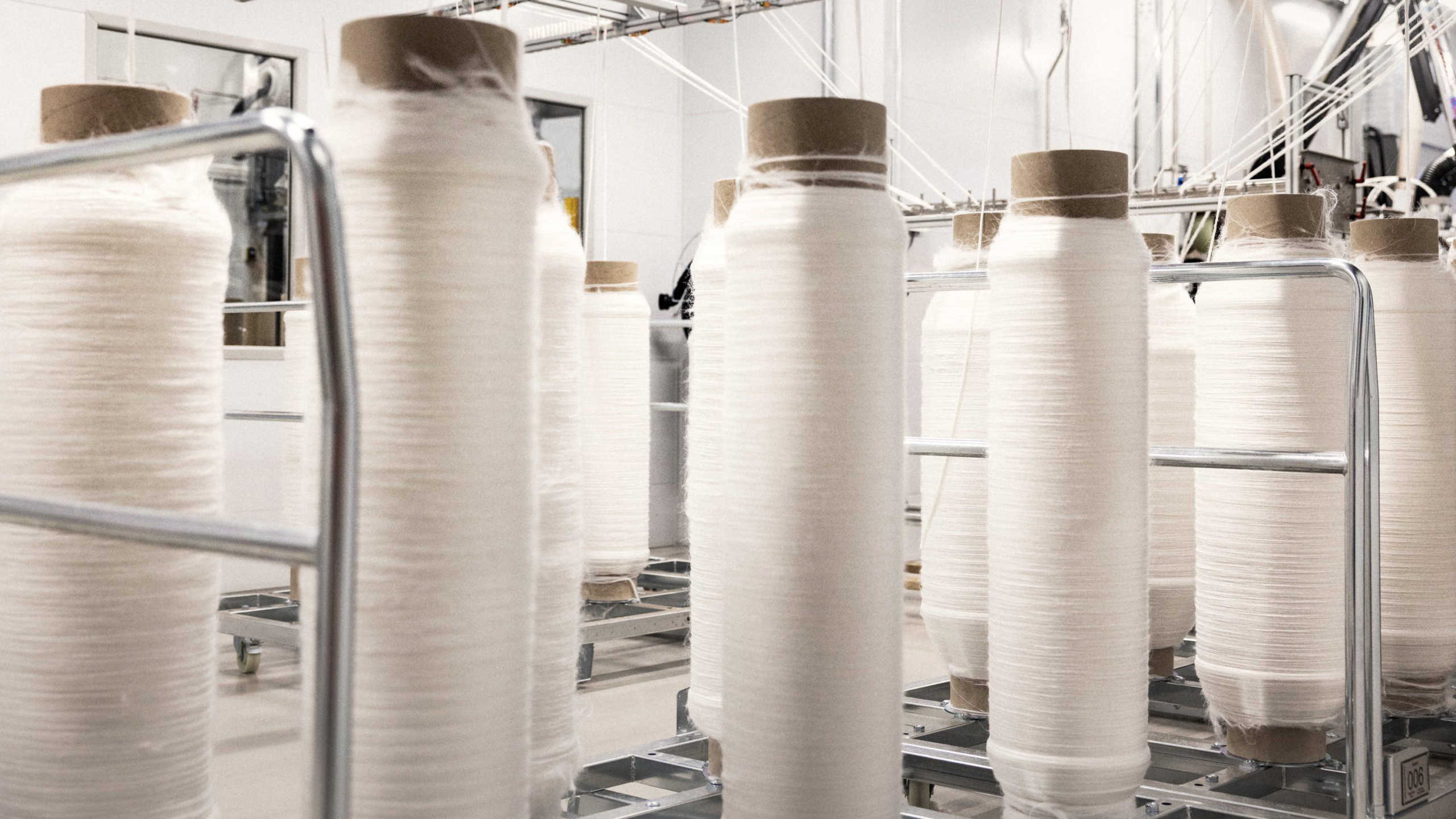
Indeed, Spinnova was able to mechanically process wood pulp into microscale fibres, which are aligned in a chain and drawn out at high pressure through a tiny nozzle into a cotton-like thread. The fibres are then dried and collected, ready to be spun into yarn. “All artificial cellulose fibres are based on dissolving processes – we do not do any dissolving of the raw material,” explains Poranen.

Janne Poranen

These new fibres use 99.5% less water and 74% less CO₂ emissions than conventional cotton, and are both recyclable and biodegradable. In addition to being chemical-free, Spinnova fibres are also free of microplastics. By 2033 the company estimates that its fibres could replace 4% of the world’s €44bn cotton supply, putting less strain on the environment and potentially improving safety for textile workers.

It took Poranen and Salmela eight years from setting up the company to commercialization, and today they have 37 international patents and more than 40 patents pending. Poranen believes that their physics background has set them up well for this long-yet-exciting journey towards solving the fashion industry’s dependence on environmentally harmful fabrics.

Going with the flow

Poranen’s pathway as a physicist began in unusual circumstances. He had just finished his compulsory military service in Finland, when he came home to find a letter from one of the many universities he had applied to. It said Poranen could study physics without taking an entrance exam because he had got such good marks at high school. Poranen duly started at the University of Jyväskylä in Finland, from which he graduated in 1997, along with a teaching degree. “I was – and still am – going with the flow,” he says.

Clouds of cellulose

Poranen had initially planned to become a physics teacher, but after really enjoying his Masters in flow dynamics and rheology at the same university, in conjunction with the pulp and paper company Valmet, he decided to continue with a PhD. “At the beginning I didn’t have any plans to do a PhD because I thought physics was way too hard for me,” says Poranen. “I was selected for a graduate programme that had close cooperation between industry and the university, and this was where I looked at what kind of rheological properties are needed to get certain kinds of paper-coating applications.”

Poranen also worked at VTT as a research scientist during his PhD, and before completing it in 2001, ended up working as an exchange researcher for more than a year with Douglas Bousfield at the University of Maine in the US. There, Poranen learnt more about developing simplified models to represent industrial processes, such as paper coating and printing, and how to verify these with experiments. When paper fibres are mechanically treated, fine-scale fibrils called cellulose nanofibrils (CNF) are generated. As Poranen learnt, these can be used in various applications such as coating, paints and medical devices. Later, Bousfield ended up as Poranen’s PhD examiner.

Infographic showing the environmental impact of textiles

Early on, Poranen knew that he preferred applied physics, and that a traditional academic career was not the path for him. After his PhD, he continued as a research scientist at VTT, but soon took on managerial roles in the forestry sector, which essentially involves overseeing products, activities and the management of forests and woodlands – be it timber, wildlife studies, biodiversity or recreation. Poranen covered a variety of jobs, from building new customer consortiums to managing R&D studies from six to eight teams as a technology manager. “Our team’s main achievement was to develop VTT’s research in the forest sector into globally leading research,” says Poranen, who remained in that position for nearly eight years before moving to work on VTT’s research in biomaterials. He became head of this department, managing six research teams and 120 employees.

In 2011 VTT was selecting its future leaders to attend a year-long virtual business-management and innovation programme at IMD Business School in Lausanne. Poranen was delighted to be one of the few who was chosen to attend, from a pool of more than 3000 researchers. Indeed, he believes it was this programme that gave him the confidence to set up Spinnova. This was where he “learned about leadership, management and strategy, along with helping me to look at the bigger picture. And it made me – someone from the central Finland forest – bold enough to come up with radical innovations.”

Spinning out

Although Poranen had been head of biomaterials at VTT for around a year, he knew in his gut that the technology being developed by Salmela’s team was revolutionary. “It was the best innovation that I had ever seen coming out of the pulp and paper sector,” he says. While Poranen was not personally involved in the details of the technology, he found that “with my physics background, it was easy for me to understand that this was a radical innovation that had to be taken to industrial scale”.

Spinnova fibre

Poranen had met Salmela during his undergraduate studies in physics. By combining their expertise, he believed that together they could move their patented idea of producing textile fibre out of cellulose forward, and transform the fashion industry. Three key researchers from Salmela’s team – two physicists and one engineer – joined them from the start, making it easier for the company to take off. “We had all the competence we needed but had to come up with ways to scale the technology from the lab; there was a lot of trial and error,” says Poranen. Today, Salmela is Spinnova’s chief technology officer.

At first, Spinnova looked at making filament yarn from paper–pulp fibres, but ditched that for sustainable microfibrillated cellulose (often referred to as nanocellulose) two years later, after buying the intellectual property from VTT. “That was a big decision,” says Poranen. Despite changing roles – from head of biomaterials at VTT to chief executive of Spinnova (a position he held until 2022) – not much changed, as he continued to be involved with the strategy and funding. But Poranen acknowledges that working for his own company made his responsibilities and workload larger. “I was working 24/7 for the last seven years,” says Poranen.

Fibre spinning wheels

But all his hard work paid off. In 2019 Spinnova finally started a pilot-scale production facility in Jyväskylä, Finland. Several renowned clothing brands such as H&M, Adidas and Marimekko had already taken an interest in the fibre and started working with Spinnova’s research and development. Its initial production facility was just a basement, but “in 2021 we were able to convince ourselves and our partners, Brazil-based Suzano – the world’s largest hardwood pulp producer – that we were ready to scale this up into the commercial level,” he says, adding that the process only makes “something like one tiny hair” so it has been a challenge to scale up.

As it happens, at the end of May this year, the first commercial facility producing SPINNOVA® fibre was launched. Operated by Woodspin – a joint venture between Spinnova and Suzano – the aim is to annually produce 1000 tonnes of its textile fibre from responsibly grown eucalyptus trees. “Spinnova’s patented fibre production process doesn’t require any harmful chemicals or dissolving, nor does it generate waste or microplastics,” explains Salmela. He adds that the process “has a 74% smaller life cycle carbon footprint and uses 99.5% less water compared to conventional cotton production. The result is a natural, cotton-like textile fibre that meets the rigorous environmental and performance demands of brands and consumers alike – and, through facilities such as this one, can now be produced at scale.”

Scaling globally

Although Poranen is no longer responsible for Spinnova on an operational level, he is excited that the company has finally made it to commercial scale. In his new role as executive chairperson of Spinnova’s board, he can now step back and look at the company’s long-term future. “As CEO I was basically responsible for everything; everybody is always coming back to you, asking if this or that is okay to do or how to move forward, and so on,” he explains. “You are practically running the whole company and it is an emotional rollercoaster because one day the company can be in a bad position but fine the next.”

Models wearing anoraks in a forest

Poranen’s goal is for Spinnova to become a world-leading sustainable textile fibre company. “The big dream is that we are able to scale it up globally,” he says. For Poranen, readily available wood pulp is Spinnova’s best bet for achieving mass scale. But, in principle, Spinnova’s technology can use any type of cellulose – be it agricultural or biowaste-based cellulose, or leather and textile waste – to produce its fibre. In fact, as of September this year, Spinnova partnered with Swedish textile-recycling company Renewcell to spin textile-waste-based fibre into new, bio-based textile fibre. Renewcell’s technology allows it to recycle textile waste such as cotton and viscose into a biodegradable pulp product called “Circulose”, which can then be used to produce new fibre. So far, Circulose has only been used to create artificial cellulosic fibres, such as viscose. By partnering with Spinnova, Circulose pulp can now be used to fabricate bio-based textile fibre, without the use of any harmful chemicals in the fibre-spinning process. Indeed, Spinnova has already produced the first batches for yarn and fabric using 100% Circulose, and made the first prototypes from a blend of cotton and Circulose-based Spinnova fibre. Spinnova estimates that the first consumer products will be available by the end of 2024.

Despite the challenges, Poranen believes that his physics expertise has prepared him for everything. “Physics itself is extremely tough, but you learn how to solve almost impossible problems,” he says. “The biggest lesson physics has taught me is to not be afraid of whatever challenges come your way and to keep moving forward.”

Pierre Agostini, Ferenc Krausz and Anne L’Huillier win 2023 Nobel Prize for Physics

Pierre Agostini, Ferenc Krausz and Anne L’Huillier have won the 2023 Nobel Prize for Physics for “experimental methods that generate attosecond pulses of light for the study of electron dynamics in matter”.

L’Huillier is based at Sweden’s Lund University; Krausz is at the Max Planck Institute of Quantum Optics and the Ludwig-Maximillians University of Munich in Germany; and Agostini is at Ohio State University in the US. The prize is worth 11 million kronor (one million dollars) and is split equally between the winners. The prize will be presented in Stockholm on 10 December 2023.

Speaking by phone at the prize announcement, L’Huillier said she was teaching when she received the news and only answered after the third or fourth call. “The last half hour of my lecture was a bit difficult to do…For me teaching is very very important!”

L’Huillier added, “This prize really means a lot. It’s the most prestigious prize and I’m so happy to get it. It’s incredible. There are not so many women who get this prize so it’s very special.”

Difficult experiments

She also spoke about the importance of fundamental research, saying that the work done by the three laureates is only now being used in practical applications after many years of development. She added that the Holy Grail of the field is to be able to control aspects of important chemical reactions such as photosynthesis. “I was personally fascinated by this field from the start, which is why I continued with it for many, many years. But it’s a slow field with difficult experiments.”

Speaking after the award was announced, Nobel Committee for Physics member Mats Larsson explained the significance of the trio’s research in terms of how it has boosted our understanding of the behaviour of electrons in chemistry and materials science.

“To me, I think the most exciting thing is that, over a very long period, [the laureates] have been working very hard both to understand the fundamental physics and then to develop the technology to really open the world of electrons, which are the workhorse for all of us and we never had access to earlier. That’s fantastic. We can study electron dynamics in atoms, molecules and condensed matter and understand, in a totally different way, how things work. It’s definitely a new world.”

He added that the laureates’ work “won’t affect our lives now, but will in the future, with applications in molecular fingerprinting and in the semiconductor industry. Molecular fingerprinting is about finding out if you have a change in your blood cells. If you can find these changes and identify them to be caused by disease, that’s a huge advantage”.

A life in science

Krausz was born in Mór, Hungary, in 1962. He studied theoretical physics at Eötvös Loránd University, graduating in 1985. After a stint as a researcher at Budapest University of Technology and Economics, in 1988 Krausz moved to Vienna University of Technology where he held various positions. In 2004 he moved to the Ludwig Maximilian University of Munich where he also became a director of the Max Planck Institute for Quantum Optics in Garching, Munich.

L’Huillier was born in 1958 in Paris. She completed her PhD in 1986 from the Pierre and Marie Curie University in Paris. After positions at the French Alternative Energies and Atomic Energy Commission (CEA), the Chalmers Institute of Technology in Gothenburg, Sweden, and the University of Southern California, Los Angeles, in 1995 she moved to Lund University in Sweden where she has remained since.

Agostini earned his doctoral degree from Aix-Marseille University in 1968. He then became a researcher at CEA Paris-Saclay and held positions at the University of Southern California, FOM Institute for Atomic and Molecular Physics in Amsterdam, and Brookhaven National Laboratory in the US. In 2005 he joined Ohio State University where he is currently based.

L’Huillier, Agostini and Krausz pioneered the generation of ultrashort light pulses with durations in the attosecond (10−18 s or 1 as) range. This is roughly the time scale on which electrons in atoms, molecules and materials interact to give rise to chemical reactions and the properties of materials. Attosecond pulses allow researchers to observe these processes unfold, providing profound insights into the chemical and material world. However, such short pulses are incredibly difficult to create, manipulate and detect in laboratory experiments.

Plateau of harmonics

L’Huillier’s contributions to the field began in the 1980s when she studied the high-frequency harmonics of ultraviolet light that are created when an infrared laser pulse is transmitted through noble gases. Under the right conditions, these harmonics can interfere with each other to create pulses of light. To make a very short pulse, light across a wide band of frequencies must be combined. Indeed, the shorter the pulse, the broader the bandwidth of light required to make it. L’Huillier and colleagues at CEA-Saclay discovered that higher harmonic generation (HHG) in noble gases produces a wide plateau of harmonics that could be combined to generate ultrashort ultraviolet pulses.

However, creating useful pulses that could be characterized proved to be very difficult. It was not until 2001 that Agostini managed to create a train of pulses each 250 as in duration. Also working at CEA-Saclay, his team re-combined the ultrashort ultraviolet pulses with the original infrared light to create an interference effect that allowed him to characterize the length and repetition rate of the pulses.

At around the same time, Krausz was working at the Vienna University of Technology where he and his colleagues developed a technique to separate individual ultrashort pulses from a train of pulses. These isolated pulses were 650 as long and were used to study a process in which electrons are removed from their atoms.

Ever shorter pulses

More recently it has become possible to create pulses that are just tens of attoseconds in duration. The work of Krausz, L’Huillier and Agostini has inspired a growing community of attosecond scientists to use ever shorter pulses to study the behaviour of electrons in a range of systems. Studies include measurements of how tightly electrons are bound to atoms and the oscillations of electrons in molecules and materials.

The technique is also starting to be used in more practical applications. For example, the presence of a molecule in a medical sample could someday be determined by the unique signal emitted by its electrons after being excited by ultrashort light pulses.

Theoretical physicist Kenneth Schafer from Louisiana State University has worked with both L’Huillier and Agostini since the 1990s and says he is “extremely happy” for them. “[L’Huillier] is an amazing scientist and human being and Agostini is a towering figure in both electron and photon physics the last 50 years,” Schafer told Physics World. “Agostini has time and again pushed the field forward, and this is just one of his many discoveries that is celebrated today.”

Schafer adds that all three laureates have driven the field of attosecond science forward and “committed to finding a deeper understanding of their results” so he feels that the Nobel committee has made the right choice. “[Their work] has resulted in close collaborations between experimentalists and theorists, and they are to be commended for it,” he says. “But the basic discoveries are theirs alone.”

That view is backed by attosecond researcher Mauro Nisoli from the Polytechnic University of Milan, who has worked with both Krausz and L’Huillier since the 1990s. Nisoli told Physics World that he is “delighted” that the field has been recognized and the recognition for the trio is “well deserved”.

“Attosecond science has become a very active field of research, with prominent applications in atomic, molecular and solid-state physics,” adds Nisoli. “Agostini, L’Huillier and Krausz carried out pioneering and crucial experiments, which led to the development of the attosecond research field.”

Oxford Instruments logo

Physics World‘s Nobel prize coverage is supported by Oxford Instruments Nanoscience, a leading supplier of research tools for the development of quantum technologies, advanced materials and nanoscale devices. Visit nanoscience.oxinst.com to find out more.

Tiny vibrating cavity sees mid-infrared light at room temperature

A new, streamlined method of “seeing” vibrations in molecules could have applications in real-time gas sensing, medical imaging, astronomical surveys and even quantum computing. These molecular vibrations occur in the mid-infrared (MIR) range of the electromagnetic spectrum, and the standard way of observing them requires detectors to be cooled to minimize thermal noise due to random high-frequency vibrations of atomic bonds. However, a team of researchers from the universities of Birmingham and Cambridge, UK have now found a way around this requirement by converting low-energy MIR photons into high-energy visible photons.

“Our new ability to see vibrations in individual molecules at room temperature, not possible before, stands out, particularly since such vibrations are typically obscured by thermal noise,” explains the Cambridge nanoscientist Jeremy Baumberg, who led the research effort. According to Rohit Chikkaraddy, a physicist at Birmingham and the first author of a Nature Photonics paper about the technology, the new method could shed light on interactions between lipids and proteins within cells, which are pivotal for understanding cellular functions that depend on molecular vibrations in the MIR range. “Our results pave the way for understanding such molecular dynamics,” Chikkaraddy says.

MIRVAL

In the new method, known as Mid-Infrared Vibrationally-Assisted Luminescence (MIRVAL), the researchers assembled molecules that emit light in the visible range into a photonic structure known as a nanoplasmonic cavity that resonates in both visible and MIR wavelength ranges. This nanoplasmonic cavity is key to the method’s success, Chikkaraddy tells Physics World. “These ultrasmall light-trapping cavities, formed by single-gold-atom defects on metallic defects, allow us to confine visible light into extremely small volumes of less than 1 nm3 and MIR light all the way down to the scale of a single molecule,” he explains.

The team then further engineered this cavity so that the vibrational states of the molecules (which absorb MIR light) and their electronic states (which absorb visible light) were able to interact. “When our system is exposed to visible light with photon energies below the light absorption electronic band, we don’t see any luminescence,” notes Baumberg. “However, when we introduce MIR light as well, the combination of visible and MIR light is sufficient for joint excitation of the molecules to occur which results in visible luminescence.”

In this way, the researchers can upconvert the low-energy MIR light to visible light, allowing them to detect the MIR light, using, for example, advanced silicon cameras like those found in smartphones.

Bringing together three vastly different length scales

One unusual aspect of the technique is that it combines the physics of three different length scales in a single platform. “These are visible wavelengths (of hundreds of nanometres), molecular vibrations (less than a nanometre) and the MIR range (ten thousand nanometres),” Chikkaraddy says.

In applications terms, the technique should make it simpler to record the vibrational “fingerprints” of individual molecules at MIR frequencies, he adds. “Through further work this novel method could not only find its way into practical devices that will shape the future of MIR technologies but also unlock the ability to coherently manipulate the intricate interplay of atoms and bonds in molecular quantum systems,” Chikkaraddy says.

The Birmingham-Cambridge researchers say they would now like to apply their technique to more complex systems, including biological entities such as lipid membranes. “This would allow us to observe the molecular dynamics of life in this new spectroscopic window,” says Chikkaraddy.

Why is there no Nobel physics prize for nuclear fission?

This summer, the theoretical physicist Robert Oppenheimer captured widespread public fascination thanks to the film Oppenheimer. The unlikely blockbuster details the life of Oppenheimer as he led the development of the atomic bomb and the conflict he couldn’t escape after it was dropped.

Tomorrow, the winner(s) of the 2023 Nobel Prize for Physics will be announced and that has led me to wonder why Oppenheimer never won a prize (he was nominated three times). Indeed, a further review of the Nobel prize archives reveals an even more puzzling question: why has there never been a physics Nobel given for the discovery of nuclear fission? Surely, the discovery of such a phenomenon warrants this recognition.

The answer lies in the stories of Otto Hahn and Lise Meitner, a German radiochemist and Austrian–Swedish nuclear physicist, respectively, who lived and worked through the rise of Nazism and the Second World War.

Complementary colleagues

The pair worked closely throughout the first decades of the 20th century, isolating and studying radioactive nuclei. Their complementary working relationship shone in the years following Enrico Fermi’s discovery in 1934 that neutron bombardment could be used to transform one element into another – work that earned Fermi the 1938 physics Nobel.

However, in 1938 Meitner – an ethnically Jewish woman – was forced to flee Nazi Germany and escaped to Sweden. There she worked at a facility that had just been set up by the physics Nobel laureate Manne Siegbahn. Meitner did not have funding to do research in wartime Sweden and some scholars suggest that her presence was resented by Siegbahn, and that he attempted to block her professional career in Sweden.

Meanwhile in Germany, Hahn continued his work along with a new assistant Fritz Strassman. During that year, Hahn and Strassman consulted Meitner on the puzzling products of the neutron bombardment of uranium, which resulted in an element too light to be the product of any known radioactive decay process. Stories say that on Christmas Eve 1938, Meitner and her nephew Otto Frisch (who would later work on the Manhattan Project with Oppenheimer) concluded that the uranium atom had been split.

Distinct papers

Hahn and Strassman published their experimental findings in the journal Naturwissenschaften in January the next year. Meitner and Frisch published their theoretical interpretation of these findings separately in Nature just a couple weeks later. These papers were distinct to their disciplines: chemistry and physics respectively.

In this situation, who would you say discovered nuclear fission – the people who performed the experiment or the people who interpreted its results? Some scholars suggest that Hahn was keen to frame the discovery of nuclear fission as a chemical discovery, not a physical one. Perhaps this was out of self-preservation, as collaboration with a Jewish woman would have ruined Hahn’s career in Nazi Germany. Fission might protect Hahn and his laboratory during those tumultuous times.

In 1944 it was announced that the Nobel Prize for Chemistry would be awarded to Hahn for the discovery of nuclear fission and no-one else, notably excluding Meitner, Strassman and Frisch. Prominent physicists, including Niels Bohr, Max Planck and Arthur Compton, protested Meitner’s exclusion and nominated her for the physics prize in the years that followed but to no avail.

So why was there no Nobel Prize for Physics awarded for fission in the subsequent years? In the case of Meitner, some scholars suggest that Siegbahn’s animosity prevented her from winning. As a leading Swedish physicist, he would have held considerable sway over who was given the prize.

And then there are the atomic bombs that were dropped on Japan in 1945, killing as many as 226,000 people. Both of these devices relied on nuclear fission, which was seen in an increasingly negative light as the threat of all-out nuclear war became real in 1949, when the Soviet Union tested it first bomb.

In the 1950s several German nuclear scientists, including Hahn, signed the Gottingen Manifesto declaring they would not participate in arming West Germany with nuclear weapons. They were ready to move past the subject, and perhaps the Nobel committee was ready to do the same.

Oxford Instruments logo

Physics World‘s Nobel prize coverage is supported by Oxford Instruments Nanoscience, a leading supplier of research tools for the development of quantum technologies, advanced materials and nanoscale devices. Visit nanoscience.oxinst.com to find out more.

Collaborative product innovation: mapping the A to Z of the SGRT clinical opportunity

The LUNA 3D surface-guided radiotherapy (SGRT) system is designed to provide end-to-end patient positioning and monitoring from CT simulation through to the treatment delivery for bore-type and C-arm linacs. Developed by German laser and radiotherapy QA specialist LAP, and officially unveiled this week at the ASTRO Annual Meeting in San Diego, California, LUNA 3D exploits high-resolution CMOS stereoscopic cameras to support precise, dose-free patient set-up and monitoring for advanced radiotherapy modalities, ensuring that the patient remains in the planned position for the duration of the treatment workflow.

For context, SGRT represents a significant diversification and evolution of LAP’s existing healthcare portfolio. That product offering, to date, spans patient positioning (for efficient surface marking of tumour position during CT/MR imaging and correct patient set-up ahead of treatment); RadCalc software for dosimetric verification in patient QA; a range of hardware phantoms to support MR imaging and radiotherapy QA; as well as multileaf collimators for precise beam-shaping and targeted tumour irradiation.

Thomas Speck

“What’s just as significant,” explains Thomas Speck, LAP’s vice-president of new product solutions (healthcare), “is that LUNA 3D leans into – and directly benefits from – LAP’s accumulated domain knowledge in laser projection and laser metrology serving industries as diverse as steel production, composite processing and concrete parts manufacturing.”

With all those dots to join, the move into the SGRT market represents one of LAP’s most ambitious product development initiatives yet. “Alongside all the technology innovation that’s gone into LUNA 3D,” notes Speck, “we have implemented a far-reaching transformation programme across core business functions – including sales and marketing, technical support and training, also our early-stage engagement with the clinical community and ‘lighthouse customers’. That realignment will ensure we are positioned to take full advantage of the commercial and clinical opportunities in SGRT.”

Delivering the clinical upside

Operationally, the LUNA 3D value proposition is all about ease-of-use for the radiation oncology team. That means intuitive and flexible browser-based user interfaces; pre-defined “treatment steps” for quick and easy selection of SGRT parameters; and synchronized data presentation across in-room displays (including tablet devices) and control-room workstations. Workflow support is also designed to be as friction-free as possible with a virtual laser for fast and easy patient set-up; automated import of patient and treatment data; and all SGRT data easily accessible by clinical staff for preparation, reporting and decision-making throughout the treatment cycle.

“Automation is hard-wired across the LUNA 3D workflow,” says Speck. “That’s because we simply can’t countenance any extension to the allocated treatment slots or additional requirement for specialized staffing. In this way, SGRT takes things to the next level, yielding new capabilities in that niche between laser-based patient positioning and patient QA.”

With more data points to guide patient set-up and alignment on the treatment couch, the expectation is that LUNA 3D will help clinical teams to fast-track the continuous improvement of their processes and treatment outcomes. At the same time, SGRT supports the online monitoring of patient position during treatment – a must-have for advanced treatment techniques like deep-inspiration breath-hold radiotherapy (which maximizes separation between the heart and lungs during radiation delivery to the latter) or stereotactic radiosurgery with couch rotation (for delivery of high-dose single fractions to small, precisely located tumour volumes).

Another notable feature of LUNA 3D is the system’s built-in scalability – essentially an SGRT solution that can be tailored to a range of user budgets for new CT and linac installations or upgrades (and without adversely impacting the time taken for linac installation, commissioning and QA). All of which is reinforced by a comprehensive SGRT training package, with classroom training to address workflow basics followed by hands-on experience and go-live support in the treatment room. After commissioning and acceptance, for example, it is envisaged that LAP application specialists will be in attendance while the clinic puts the LUNA 3D system through its paces on the initial cohort of patients.

Collaborative innovation

Zooming in on product development and technology innovation, it’s evident that LUNA 3D is very much the outcome of a collective and cross-disciplinary effort. “Throughout the journey from project initiation to finalized SGRT offering,” says Speck, “LAP’s R&D and product engineering teams worked hand-in-hand with a network of clinical partners at the sharp-end of treatment delivery. Our goal was to understand – in granular detail – how to design an SGRT clinical workflow that’s as easy to implement as the workflow for a positioning laser.”

It helps, in this regard, that LAP can tap into an established global consortium of clinical partners, with each site offering varying levels of engagement – whether that’s clinical consultancy, participation in usability studies, or beta-testing of the early-stage SGRT product demonstrator in a preclinical setting. As a result, clinical feedback “from the field” informs all aspects of the LUNA 3D product design: from the hardware and optical profiling capabilities through software development, automated workflows, as well as the implementation of open data interfaces to ensure seamless integration of SGRT with other systems in the treatment room.

Right now, for example, LUNA 3D is being evaluated at several early-adopter sites around the world – among them Pius Hospital in Oldenburg, Germany; CCGM Clinique Clémentville in Montpellier, France; and Penn Medicine in Philadelphia, PA, US.

When it comes to the specifics of product roll-out, LAP is aiming to have LUNA 3D cleared for full commercial release in the US in the final quarter of this year – pending 510(k) approval from the US Food and Drug Administration (FDA) – while the CE mark on the same timeframe will provide a green light for sales to clinical customers in the European Economic Area. Speck and his colleagues have also initiated the regulatory approval processes for priority markets in Asia, including Japan, Singapore and Korea.

LAP offers different LUNA 3D service packages to meet individual customer needs including, for example, annual maintenance, software/hardware upgrades, installation and training.

SGRT made easy

LUNA 3D represents an ambitious extension to LAP’s patient positioning and QA portfolio for radiotherapy. Other key take-aways include:

  • The SGRT system works independent of patient skin tone owing to blue-coloured speckle projection.
  • Multiple displays (including tablet devices) in the treatment room ensure the clinical team has all key SGRT data in its line-of-sight (as well as remote data access via mobile devices).
  • All necessary calibration and QA tools are supplied as standard, including expandable LAP EASY CUBE phantom.
  • Site-specific installation is conducted by LAP room-planning experts and service personnel.

German hyperinflation, and what it has to do with a Nobel prize

Many of us are feeling the sting of inflation. But did you know that runaway prices could well have prevented Otto Stern from receiving a Nobel prize?

Stern was a German physicist who is best known for the Stern–Gerlach experiment, which was done in 1922 with fellow German Walther Gerlach. While the experiment was first interpreted as important evidence for quantum mechanics, the theory it was based on turned out to be wrong. However, it was still an astonishing result and today the Stern–Gerlach experiment is regarded as evidence for the intrinsic angular momentum (quantum spin) of particles such as electrons.

But, the experiment may have never happened because in 1922, hyperinflation was rampant in Germany, and Stern and Gerlach were struggling to pay for their expensive equipment. Max Born, who Stern worked for, helped out by donating money raised from his public lectures on quantum mechanics. Heeding the advice of a friend, Born also wrote to Henry Goldman, a prominent American banker and son of the founder of Goldman–Sachs. Goldman, who had actually retired from his father’s firm by this time, was a philanthropist and sent Born a cheque for “some hundreds of dollars” (somewhere around £10,000 today) that saved the experiment. Albert Einstein also donated some money to the Stern–Gerlach cause. He had been a mentor of Stern’s.

Thanks to these generous donations, the experiment was a success, but neither Stern nor Gerlach won a Nobel prize for their famous experiment. However, in 1943 Stern received the Nobel Prize for Physics “for his contribution to the development of the molecular ray method and his discovery of the magnetic moment of the proton”. Both achievements came about in part because of his efforts on the Stern–Gerlach experiment.

Oxford Instruments logo

Physics World‘s Nobel prize coverage is supported by Oxford Instruments Nanoscience, a leading supplier of research tools for the development of quantum technologies, advanced materials and nanoscale devices. Visit nanoscience.oxinst.com to find out more.

Copyright © 2026 by IOP Publishing Ltd and individual contributors