Exchange bias in a single-layer film is created using ion implantation

A new method for creating and controlling exchange bias in an antiferromagnetic single-layer film has been developed by researchers in the US and Switzerland. The method involves low-energy ion implantation into the film, and it has the potential to advance the development of antiferromagnetic spintronics devices.

Exchange bias refers to a shift in the position of the hysteresis loop of a ferromagnet along the horizontal axis (applied magnetic field). This can occur when an antiferromagnet layer and a ferromagnet layer are brought close together, pinning the magnetization of a ferromagnetic layer. Exchange bias materials play important roles in spintronic technologies such as the read head sensors in hard disk drives; spin valves; and the magnetic tunnel junctions used in digital memories.

Generating a strong exchange bias field between a ferromagnet and an antiferromagnet requires a good quality interface between the two materials. However, achieving this in thin films can be challenging. Moreover, to obtain exchange bias, the system must be heated above the antiferromagnet’s Neel temperature and then cooled in the presence of a magnetic field.  This thermal treatment can cause diffusion between the two layers, which reduces significantly the exchange bias. To prevent this, diffusion barriers are generally incorporated into the stack structure. However, this increases the complexity and thickness of the device, making it unsuitable for the miniaturization of spintronic devices.

Material for the future

Now, Cory Cress  and Steven Bennett at the US Naval Research Laboratory, and researchers there and at the Paul Scherrer Institute and Oak Ridge National Laboratory have used low-energy ion implantation to create an appreciable exchange bias in the antiferromagnetic material iron–rhodium (FeRh). And, they have done this without the need for a ferromagnetic layer.

Ion implantation is a common technique in materials science and engineering and is used to modify the properties of a material by introducing ions into its surface or bulk. The process involves creating a beam of ions that is then directed at a target material.

In their experiments, the team first deposited a single-crystal FeRh film onto a magnesium oxide substrate (see figure below). The sample was then heated to 730 °C and cooled in a process called annealing. This put the FeRh film into a face-centred cubic-type crystal structure. This phase exhibits a high-speed meta-magnetic transition from an antiferromagnetic to a ferromagnetic state when the temperature exceeds about 400 K. Additionally, the FeRh film possesses a switchable magnetic moment that can quickly flip direction, making it promising for various spintronic applications.

Exchange bias image

Taking a cue from their previous study of tuning a meta-magnetic transition temperature in FeRh through ion implantation, the researchers implanted low-energy helium or iron ions into FeRh films at room temperature. This created ferromagnetic surface layers that were adjacent to the antiferromagnet layer. This created a relatively large exchange bias effect of 41 Oe for the iron-implanted film and 36 Oe for the helium implanted film.

According to the team, these findings are promising because the exchange bias effects previously observed in FeRh have been typically very low or only seen below room temperature. As a result, the team believes creating exchange bias by implantation in FeRh could lead to the development of advanced antiferromagnetic devices.

The origin of exchange bias has remained a long-standing puzzle since its discovery, despite the development of various models that try to explain it. In this study, Cress, Bennett and colleagues aimed to shed further light on the origin of this phenomenon by subjecting ion implanted FeRh films to one hour of annealing either at 400 °C or 700 °C. After the annealing process, the team noted a substantial decrease in the exchange bias field of both implanted films. This was attributed to the healing of defects formed during implantation and restoration of the film’s antiferromagnetic state near the surface.

Defects are responsible

By using annealing to regain the pristine properties of the helium implanted film, the researchers found compelling evidence that any exchange bias present in the film is due to the defects caused by the ion implantation and not because of any changes in the FeRh composition due to the addition of iron ions. Furthermore, the team used a domain state model to explain these results, proposing that non-magnetic defects within the antiferromagnetic layer play crucial role in forming and stabilizing the exchange bias field.

To gain further insight into the exchange bias of FeRh films, the team employed polarized neutron reflectometry. This technique measures magnetization as a function of depth by directing polarized neutrons on to the surface of the film and measuring the intensity of the reflected neutrons.

Using this technique, the researchers observed the pinned uncompensated magnetic moments within the antiferromagnetic region of the film. They found that these uncompensated moments result from defects, such as vacancies created by ion implantation. When coupled with an adjacent ferromagnetic layer, the vacancies result in the exchange bias phenomenon. According to the researchers, these findings provide a convincing proof for the domain state model of exchange bias.

The team’s findings are reported in the Journal of Materials Chemistry C and could potentially lead to the application of the domain state model in diverse magnetic spin systems.

Germany reveals €3bn plan to build a quantum computer by 2026

The German government says it will spend €3bn over the next three years to build a universal quantum computer. The project is part of a new initiative to make Germany competitive with countries that have already built or are taking steps to construct such a device. It is hoped the cash will boost the German economy and place the country at the top of quantum developments in the European Union.

Set to be built by 2026, Germany’s quantum computer will exploit current quantum technology. It will have a capacity of at least 100 qubits but this could later be expanded to 500 qubits. Funding for the device includes €2.2bn split among several government ministries, including €1.37bn for the research ministry. National research institutes will receive another €800m.

The initiative also includes the commitment to build a quantum ecosystem and foster a quantum industry. Several major German companies and institutions are already active in quantum technology. The automotive supplier Bosch, for example, is working with IBM to see if quantum computing simulations could help to replace rare-earth metals in electric motors.

German laser giant Trumpf, meanwhile, is developing quantum computer chips as well as quantum sensors, while semiconductor manufacturer Infineon has developed the first quantum-encrypted computer chips. The German Aerospace Center DLR has also launched its first test satellites for quantum-key distribution.

According to German education minister Bettina Stark-Watzinger, quantum technology is  crucial for Germany’s technological sovereignty. She expects that by 2026, “at least 60 end users of quantum computing should be active in Germany”, adding that the country should “be among the top three within the EU and at least reach the level of the US or Japan [in quantum computing]”.

Giant tunnelling magnetoresistance appears in an antiferromagnet

Researchers in China have observed giant tunnelling magnetoresistance (TMR) in a magnetic tunnel junction made from the antiferromagnet CrSBr. When cooled to a temperature of 5 K, the new structure exhibited a magnetoresistance of 47,000% – higher than commercial magnetic tunnel junctions – and it retained 50% of this TMR at 130 K, which is well above the boiling point of liquid nitrogen. According to its developers, the structure can be manufactured in a way that is compatible with the magnetron sputtering process used to make conventional spintronics devices. These qualities, together with the fact that CrSBr is stable in air, make it a promising candidate platform for spintronic devices, they say.

Standard magnetic tunnel junctions (MTJs) consist of two ferromagnets separated by a non-magnetic barrier material. They are found in a host of spintronics technologies, including magnetic random-access memories, magnetic sensors and logic devices.

Junctions based on A-type van der Waals (vdW) antiferromagnets such as CrSBr and other chromium halides are an attractive alternative to conventional MTJs thanks to their unusually high tunnelling magnetoresistance. They work thanks to the spin-filter effect, in which the electron spins (or magnetic moments) of the chromium atoms in CrSBr are ferromagnetically coupled to other atoms in their layer and antiferromagnetically coupled to atoms in neighbouring layers. In other words, the spins align parallel to each other in the single layers and antiparallel to each other between neighbouring layers.

While the high tunnelling resistance of these so-called spin-filter MTJs (sf-MTJs) makes them good candidates for magnetic memories, they do have certain drawbacks. Notably, the materials they are made from tend to be unstable and prone to losing their magnetism at high temperatures. This makes it hard to use them in practical spintronic devices.

Overcoming fabrication challenges

In the latest study, researchers led by Guoqiang Yu of the Beijing National Laboratory for Condensed Matter Physics developed a new fabrication technique for these desirable materials. Working with colleagues in Beijing, Dongguan and Wuhan, they began by depositing a bilayer of platinum (Pt) and gold (Au) onto Si/SiO2 wafers using DC magnetron sputtering.

Next, members of the team mechanically shaved off thin flakes of CrSBr from a sample of the bulk material and placed them onto the Si/SiO2/Pt/Au substrates. This enabled them to obtain relatively thin CrSBr flakes on Pt/Au with clean and fresh surfaces. At this point, the researchers deposited a further layer of platinum onto the CrSBr with an ultralow sputtering power of 3–5 W and a relatively high deposition pressure of around 1 Pa. Finally, they used ultraviolet lithography and Ar ion milling to fabricate several sf-MTJs from the layered structure they created.

Promising properties

The new sf-MTJs have many favourable characteristics. “The first is that the route we employed to make them is more compatible with those employed to fabricate conventional spintronics metallic stacks,” Yu explains. “The second is that they retain 50% of their TMR even at a temperature of 130 K, which is so far the record-high working temperature for sf-MTJs.”

Yu points out that this record-high operating temperature is not far below CrSBr’s so-called Néel temperature, beyond which the material’s thermal energy prevents its spin moments from aligning. This relatively high operating temperature comes with an important practical advantage, Yu adds. “Compared to previous such junctions, our sf-MTJs might work in the liquid nitrogen temperature range and perhaps even at room temperature,” he observes. “And thanks to their stability in air, they are more suited to real-world applications.”

That is not all. CrSBr is also a semiconductor, so its neighbouring layers have opposite magnetic moments at zero or small magnetic fields. This means it can be used as a barrier layer at low temperatures. “In this configuration, all the electrons, spin-up or spin-down, must encounter a higher barrier height after being polarized in one spin direction or another by passing through the first layer because the next layer has an opposite spin orientation, giving rise to higher tunnelling resistance,” Yu tells Physics World. “When the applied magnetic field is large enough, all the magnetic moments are aligned with this field and, in this case, the electrons with spins parallel to the field direction encounter a lower barrier height, which results in lower tunnelling resistance.”

The researchers, who report their work in Chinese Physics Letters, suggest that the new junctions could be used in spintronic devices based on a stack of a just a few layers of CrSBr. “Our study has revealed that sf-MTJs based on 2D vdW A-type antiferromagnets have some outstanding properties,” Yu says. “We will now be trying to find a 2D vdW A-type ferromagnet with a higher Néel temperature to further improve the working temperature of the junction we have made so that it is more suited to applications.”

A further challenge, the researchers say, will be to figure out a way to electrically manipulate the magnetization on the A-type antiferromagnet so they can construct fully functioning spintronic devices.

Sects, drugs and drunken duels: lighter moments from the history of science

Ada Lovelace

René Descartes revolutionized philosophy, science and mathematics, but did you know he moved to Amsterdam in the early 17th century for the same reason young people continue to go there today? Yes, he went to smoke a lot of weed and get away from his father. And then he became a fanatical supporter of a weird religious sect that didn’t actually exist.

Meanwhile, Ada Lovelace – mathematical prodigy and author of the first computer program in the 1840s – was a compulsive gambler. She lost so big she sold the family jewels, and when her mother-in-law bought them back, Lovelace lost them all over again. In fact, she was still in debt when she died.

These are just two examples from The Limits of Genius: the Surprising Stupidity of the World’s Greatest Minds, written by science journalist Katie Spalding. In this informative, funny book, Spalding profiles people widely considered to have been geniuses. In each case she briefly sketches their background before digging deep into examples of when they were…not so clever.

Spalding’s style is chatty and irreverent, with quite a bit of swearing, so that at times this book reads like a series of Twitter threads – admittedly impeccably researched and heavily footnoted Twitter threads. And though you may already be familiar with some stories – such as astronomer Tycho Brahe’s penchant for getting drunk and fighting duels – the mix of people covered means there is bound to be something new to you.

As Spalding’s introduction admits, however, there aren’t many women in this book. This is largely because the women who have managed to achieve renown tend to be written about so little that their interesting quirks and flaws just weren’t recorded, which means the chapters about women feel lighter on detail.

“Stupid” is also a subjective label. Spalding is quick to point out the racism, sexism, ableism and other forms of bigotry her subjects suffered from or were guilty of. But she also includes issues that could be seen as out of an individual’s control rather than “stupid”. For example, psychologist Sigmund Freud probably didn’t know cocaine was massively addictive before he started using (and prescribing) it in huge quantities. And civil rights activist and author Maya Angelou certainly couldn’t help having a murderously dangerous mother.

On the other hand, in some cases the acts of stupidity are intrinsically linked to the scientific research being conducted. Physicist Marie Curie did carry around radioactive materials in her pockets, leading to horrible skin lesions and her early death – but it’s also in part how she figured out radioactivity. The meteorologist and aeronaut James Glaisher did nearly kill himself by taking multiple hot-air balloon flights so high that he passed out (the exact height is unknown because all his instruments broke). But from these accidents he figured out details of the Earth’s atmosphere that revolutionized the nascent field of meteorology.

So The Limits of Genius might be best described as a highly entertaining whistle-stop tour of lesser-known facts and anecdotes about well-known people. With swears.

  • 2023 Hachette 352pp £22hb
  • Sold in the US with the title Edison’s Ghosts: the Untold Weirdness of History’s Greatest Geniuses (2023 Hachette 352pp $29hb)

Ultrathin e-tattoo provides continuous heart monitoring

Cardiovascular disease is the leading cause of death worldwide. Continuous cardiac monitoring could allow earlier detection of heart disease, enabling timely intervention to prevent serious cardiac complications. Traditional monitoring devices, however, are designed for clinical settings and are too heavy and power-hungry for long-term measurements of people on the move.

A team headed up at The University of Texas at Austin aims to solve this problem with the creation of an ultrathin (200 µm) and lightweight (2.5 g) device that provides continuous cardiac monitoring outside of the clinic. The stretchable electronic tattoo, or e-tattoo, attaches to the chest via a medical dressing. It boasts ultralow power consumption (less than 3 mW), runs on a small battery with a life of more than 40 h, and wirelessly streams real-time data to a host device such as a mobile phone.

“Most heart conditions are not very obvious. The damage is being done in the background and we don’t even know it,” explains lead author Nanshu Lu in a press statement. “If we can have continuous, mobile monitoring at home, then we can do early diagnosis and treatment, and if that can be done, 80% of heart disease can be prevented.”

Dual-mode electro-mechanical sensing

The e-tattoo works by measuring two key cardiac signals: the electrical activity of the heart via electrocardiography (ECG); and mechanical cardiac rhythm (subtle vibrations caused by heart contraction and blood movement) via seismocardiography (SCG). The ECG sensor interfaces with the body using bio-compatible graphite film electrodes, while the SCG is recorded by a high-resolution, low-noise accelerometer.

Synchronization between the ECG and SCG signals enables the measurement of key cardiac time intervals – the pre-ejection period (PEP) and the left ventricular ejection time (LVET) – with high accuracy. Such time intervals are important indicators of many cardiovascular diseases, but currently can only be measured via invasive means.

“Those two measurements, electrical and mechanical, together can provide a much more comprehensive and complete picture of what’s happening with the heart,” says Lu. “There are many more heart characteristics that could be extracted out of the two synchronously measured signals in a non-invasive manner.”

An e-tattoo designed for long-term wear must be comfortable, conform to the contours of the chest and stretch with the skin as the user moves. To achieve this, the team used serpentine interconnects between the sensors and electronic circuits. They found that the e-tattoo could stretch up to 20% without any damage or drop in signal quality.

<strong>Flexible and comfortable</strong> The e-tattoo uses stretchable interconnections to conform to the body. (Courtesy: The University of Texas at Austin)

Performance comparisons

To validate the quality of the acquired signals, the researchers compared data from the e-tattoo’s sensors against gold-standard clinical devices, observing that both devices captured equivalent signals and data. Next, they tested the e-tattoo on five healthy volunteers, who wore the e-tattoo while holding static poses and cycling under incremental load with breaks. For comparison, participants also wore a non-invasive cardiac output monitor (NICOM).

During the static poses, heart rates measured by the e-tattoo and the NICOM agreed well for all participants, with a difference of 0.07±1.21 beats per minute (bpm). As participants transitioned from lying to sitting upright and then to standing, the e-tattoo recorded increased PEP and decreased LVET, as expected. This demonstrates the device’s ability to measure small changes in cardiac time intervals caused by posture variations.

In the cycling experiment, heart rate measurements from the e-tattoo and the NICOM were again highly correlated, with a difference of 0.02±1.24 bpm. The ECG signal remained pristine during cycling, but the SCG signal was corrupted by motion artefacts. Thus the researchers examined the rest periods between each cycling segment, during which the mean difference in LVET between the e-tattoo and the NICOM was −0.44 ± 8.74 ms. This linear relationship shows that the e-tattoo can provide a viable alternative to bulky and expensive clinical monitors.

Finally, the team performed a long-term wearability test with a single subject, who wore the e-tattoo for more than 24 h to demonstrate its use in day-to-day settings. The e-tattoo demonstrated good correlation with a consumer smartwatch in heart rate measurements. Manual inspection of the long-term data showed that during restful segments (such as working at a computer, pausing during a walk or sleeping), the ECG and SCG data were mostly free of motion artefacts and suitable for extracting cardiac time intervals.

The researchers describe the e-tattoo in Advanced Electronic Materials.

Iridium Netwerk’s medical physics team sees the ‘big picture’ on transit in vivo dosimetry

Electronic portal imaging devices (EPIDs) are now widely used as dosimeters by radiation oncology clinics – both for automated pre-treatment verification (without the patient present) and for transit in vivo dosimetry (with the patient in situ on the treatment couch). In the case of the latter, the motivation is to enhance patient safety by detecting errors and deviations in dose delivery (owing to changes in patient anatomy, for example) over the course of radiation treatment, while simultaneously addressing the increased patient QA complexity of advanced modalities such as volumetric modulated arc therapy (VMAT) and stereotactic body radiotherapy (SBRT).

Beyond the immediate upsides of transit in vivo dosimetry – chiefly, a final safety net for the healthcare team and the patient – there’s also the longer-term roadmap towards at-scale patient-specific quality assurance (PSQA). Put another way: fully automated, EPID-based transit dosimetry opens the way for medical physicists to not only detect divergence of an individual radiotherapy fraction versus the treatment plan, but to use the cumulative (and ever-growing) patient QA data set as a tool to re-evaluate and reimagine best practice within the radiation oncology workflow.

The engine-room of patient QA

A pioneer in this regard is Dirk Verellen, director of medical physics at Iridium Netwerk, a multi-site radiation oncology programme in the Greater Antwerp region of Belgium. Earlier this year, Verellen and his team published a granular analysis of a four-year PSQA data set spanning a large cohort of Iridium Netwerk cancer patients with diverse disease indications. Their findings are instructive, demonstrating systematic correlation between transit dosimetry measurements over time versus adaptations within the clinical workflow. “Our results suggest EPID in vivo dosimetry is able to assess the impact of some adaptations to the workflow and can therefore assist in continuous quality improvement of patient treatment and outcomes,” explains Verellen.

Dirk Verellen

At the heart of Iridium Netwerk’s PSQA work programme is the SunCHECK Quality Management Platform from Sun Nuclear (a Mirion Medical company), the US-based manufacturer of independent QA solutions for radiotherapy facilities and diagnostic imaging providers. Deployed across the Antwerp healthcare system’s four treatment centres through late 2017 and early 2018, SunCHECK comprises a single interface and database offering a unified view of patient and machine QA that’s independent from the treatment system. As such, SunCHECK’s two core software modules – SunCHECK Patient and SunCHECK Machine – are now established as the QA “engine-room” for Iridium Netwerk’s distributed medical physics service.

That service, staffed by 19 medical physicists and seven physics assistants, is built around a unified suite of Varian treatment systems (currently nine TrueBeam machines and one Clinac iX) delivering leading-edge cancer care to around 6000 patients every year. Two treatment planning systems (TPS) deal with the heavy lifting in advance of treatment delivery: RayStation (from RaySearch Laboratories, Sweden) for stereotactic plans and Varian’s Eclipse TPS for all other types of treatment plan. Meanwhile, SunCHECK Patient encompasses all aspects of Iridium Netwerk’s patient QA, including secondary checks, phantomless pre-treatment QA and automated in vivo monitoring (with the EPID-based measurements managed by SunCHECK’s dedicated PerFRACTION software module).

“With PerFRACTION, we’ve shown that large-scale clinical implementation of in vivo transit dosimetry is feasible, even for complex techniques,” says Evy Bossuyt, a senior medical physicist in Verellen’s team and project lead for the integration and ongoing development of SunCHECK Patient within the Iridium Netwerk radiotherapy programme. “In this way, PerFRACTION adds an extra dimension to patient QA, revealing a variety of deviations spanning errors in planning, machine problems, patient positioning and changes in patient anatomy such as weight loss, tumour shrinkage or rectal/bladder filling.”

Alongside the enhanced error detection, the Iridium Netwerk medical physics department has seen significant streamlining over the past five years with regards to the aggregate workload and staff-time allocated to essential patient QA checks. “That’s down to SunCHECK Patient’s high degree of automation plus the in-built accessibility that comes from a web-based software platform,” notes Bossuyt.

Data-driven insights

That emphasis on automation and online access is, by extension, fundamental to Iridium Netwerk’s retrospective PSQA study – a longitudinal review that aggregates data from all the group’s radiotherapy patients treated between September 2018 and August 2022. In total, Bossuyt and colleagues analysed 84,100 transit in vivo dosimetry measurements, dividing them into four yearly periods. The team also classified failed measurements by pathology and into four categories of failure: technical, planning and positioning problems as well as anatomical changes in the patient.

“We investigated if the observed trends in the in vivo dosimetry results versus time could be a result of adaptations to the clinical workflow,” explains Bossuyt. “Also the other way around: if the impact of adaptations could be monitored via the in vivo dosimetry.”

Overall, Bossuyt and the project team found that the number of failed measurements linked to patient-related problems gradually decreased from 9.5% to 5.6% over the four-year study period (see “Further reading”). What’s more, a deep-dive into the transit dosimetry data set reveals no shortage of success stories reflecting the impact of targeted workflow changes.

Failed measurements attributed to positioning problems, for example, decreased from 10.0% to 4.9% in boost breast-cancer patients after the introduction of extra imaging; from 9.1% to 3.9% in head-and-neck patients following education of radiation therapists on positioning of patients’ shoulders; from 6.1% to 2.8% in breast-cancer patients after introduction of ultrahypofractionated breast radiotherapy with daily online pre-treatment imaging; and from 11.2% to 4.3% in extremities following introduction of immobilization with calculated couch parameters and a surface-guided radiation therapy solution. Elsewhere, following targeted patient education from dieticians, failed measurements related to anatomical changes decreased from 10.2% to 4.0% in colorectal patients and from 6.7% to 3.3% in prostate patients.

Automate and accumulate

Verellen and Bossuyt, for their part, are already thinking about next steps regarding the clinical exploitation of PerFRACTION and transit dosimetry. One use-case under investigation is the automatic triggering of offline (ultimately online) adaptation for specific disease indications – in head-and-neck patients, for example, where the anatomy adjacent to the tumour often changes only slowly during the course of treatment.

“We’re evaluating a workflow that’s able to ‘red-flag’ significant changes in patient anatomy based on transit dose measurements over the previous three or four fractions,” says Verellen. “Automation is the key to success here,” he adds. “Using the transit dose data to show that the treatment plan is gradually decreasing in quality, the organs-at-risk are reaching their safe limits, so it might be time to replan the patient. That’s where we want to go next with our patient QA platform.”

As a SunCHECK reference site, Iridium Netwerk promotes radiotherapy QA best practice using the SunCHECK Quality Management Platform. The clinical team collaborates with Sun Nuclear on its product development roadmap while serving as a regional resource for the growing European base of SunCHECK users.

Further reading

Evy Bossuyt et al. 2023 Assessing the impact of adaptations to the clinical workflow in radiotherapy using transit in vivo dosimetry (phiRO 25 100420)

  • For more information about SunCHECK, visit Sun Nuclear on booth 150 at the ESTRO Annual Congress in Vienna, Austria (12–15 May).

Threshold for X-ray flashes from lightning is identified by simulations

New insights into how X-ray flashes are produced during lightning strikes have been made by researchers in the US, France, and the Czech Republic. Using computer simulations, a team led by Victor Pasko at Penn State University showed how avalanches of electrons responsible for the flashes are triggered at a minimum threshold the electric fields produced by the precursor to lightning. This discovery could lead to the development of new techniques for producing X-rays in the lab.

Terrestrial gamma-ray flashes (TGFs) involve the emission of high-energy photons from sources within Earth’s atmosphere. While the term gamma-ray is used, most of the photons are created by the acceleration of electrons and are therefore X-rays.

These X-rays are emitted in the megaelectronvolt energy range and their creation is closely associated with lightning. Although TGFs are rare and incredibly brief, they are now regularly observed by instruments that detect gamma rays from space.

Space telescopes

“TGFs were discovered in 1994 by NASA’s Compton Gamma Ray Observatory,” Pasko explains. “Since then, many other orbital observatories have captured these high-energy events, including NASA’s Fermi Gamma-ray Space Telescope.”

Following their initial discovery, the origins of TGFs were linked to electrons that are liberated from air molecules by the intense electric fields of “lightning leaders”. These are channels of ionized air that form between a negatively charged cloud bas and the positively charged ground. As the name suggests, the creation of lightning leaders is followed shortly by lightning discharges.

Once these electrons are liberated in a lightning leader, they are accelerated by the electric field and collide with molecules to liberate more electrons. This process continues, very rapidly creating more and more electrons in what Pasko describes an “electron avalanche”.

Ionizing X-rays

As the electrons collide with molecules, some of the energy lost by the electrons is radiated in the form of X-rays. These X-rays travel in all directions – including back along the path of the electron avalanche. As a result, the X-rays can ionize more molecules upstream from the avalanche, liberating more electrons and making the TGFs even brighter.

After this initial model was conceived in the early 2000s, researchers attempted to recreate the behaviour in computer simulations. So far, however, these simulations have not managed to closely mimic the sizes of TGFs observed in real lightning strikes.

Pasko and colleagues believe that this lack of success is related to the relatively large size of these simulations, which usually model regions that are several kilometres across. However, this latest work suggests that TGFs typically form in highly compact regions (ranging from 10 to 100 m in size) surrounding the tips of lightning leaders. Until now, the reasons surrounding this compactness have largely remained a mystery.

Minimum threshold

In their study, the researchers assumed that TGFs only form when the strength of the lightning leader’s electric field exceeds a minimum threshold value. By simulating more compact regions of space, Pasko and colleagues were able to identify this threshold. What is more, the TGFs produced in this way matched real observations far more closely than previous simulations.

Pasko and colleagues hope that future simulations could mimic the TGF electron avalanche mechanism far more closely – potentially leading to new techniques for producing X-rays in the lab. “In the presence of electrodes, the same amplification mechanism and X-ray production may involve generation of runaway electrons from the cathode material,” Pasko explains.

Ultimately, this could lead to deeper insights into how X-rays can be produced through controlled electrical discharges in gases. This could lead to compact, highly efficient X-ray sources. Pasko concludes, “We anticipate a lot of new and interesting research to explore different electrode materials, as well as gas pressure regimes and compositions that would lead to enhanced X-ray production from small discharge volumes.”

The work is described in Geophysical Research Letters.

New transport measurement system from Oxford Instruments

In this video filmed at the 2023 March Meeting of the American Physical Society in Las Vegas, Matt Martin, managing director of Oxford Instruments Nanoscience, introduces a new transport measurement system, which has been produced to allow complete integration with Lake Shore products. It uses open-source technologies, such as QCoDeS and Jupyter Notebooks, so that it can be easily tailored to meet the needs of the user and can be flexibly integrated with all third-party electronics.

As Martin explains, the user ends up with a complete data set, which they can then use for the research papers that they want to write. Using Grafana and the Jupyter Notebook tools, graphs can be created and dropped directly into an article.

Also featured in the video is Abi Graham, a measurement scientist from the company, who talks about the firm’s cryostat control software, which lets users monitor the status of their fridge and also set and change parameters such as field and temperature. This software is linked to the Proteox control unit, with the measurement server hosting a browser-based Jupyter Notebook to give full remote access.

Martin then describes how the integration with Lake Shore products was achieved. He explains that with OI:DECS and an open-framework environment, the company is attempting to create a community using the framework of Oxford Instruments software and databasing.

Multispectral infrared imaging improves guidance of cancer surgery

Enhancing tumour delineation during surgery

Surgical removal of a tumour is one of the most common treatments for cancer. To help distinguish tumours from healthy tissue during excision, surgeons use fluorescence-guided surgery (FGS) to improve the visibility of structures that may not be seen clearly in the white light of an operating room. A London-based research team has now shown that multispectral short-wave infrared (SWIR) imaging combined with machine learning shows promise to improve the accuracy of FGS.

FGS uses tumour-targeted imaging agents to provide real-time visualization of tumours with molecular specificity and high-contrast margin delineation. The target tissue is stained with a near-infrared-emitting dye that preferentially binds to the surface of tumour cells. Most FGS set-ups use the absolute intensity of the infrared emission to discern which image pixels correspond to tumours. This intensity, however, is sensitive to the lighting conditions in the operating room, the camera setup, the amount of dye used and the time elapsed after staining. As such, intensity-based classification is prone to inaccuracies.

Dale Waterhouse

Writing in the Journal of Biomedical Optics, the team explains that by capturing multispectral SWIR images of the dyed tissue, and using machine learning to classify pixels based on their spectral characteristics, rather than intensity, more robust delineation of tumour tissue during FGS is possible.

Led by Dale Waterhouse of University College London, researchers from the UCL Great Ormond Street Institute of Child Health and the Great Ormond Street Hospital for Children NHS Foundation Trust created a multispectral SWIR fluorescence imaging system. The system illuminates tissue with a 785 nm fibre-coupled laser and uses a highly sensitive InGaAs camera coupled to a SWIR lens to collect the fluorescence emission. This fluorescence light is sequentially filtered through six long-pass filters, with cut-off wavelengths of 850, 950, 1050, 1150, 1250 and 1350 nm.

The researchers assessed their system using an animal model of an aggressive type of neuroblastoma, following injection of a neuroblastoma-specific fluorescent probe. They sequentially placed each filter in front of the SWIR optical system and captured six images, using these to construct image cubes (640 × 512 pixels × six filters) representing the fluorescence collected from 850 to 1450 nm.

Next, the team trained seven machine learning-based models to classify each pixel as tumour, non-tumour tissue or background, based on their spectra. The researchers also compared different normalization approaches designed to make pixel classification independent of the absolute intensity.

Using AUC=1 spectra normalization, the best performing model achieved a per-pixel classification accuracy of 97.5% (97.1%, 93.5% and 99.2% for tumour, non-tumour tissue and background, respectively). The team note that the normalization strategy made the results of the model far more robust to changes in imaging conditions.

Looking to the future

Waterhouse and collaborators advise that a second-generation system will require another method of multispectral imaging with higher temporal resolution, because image cube acquisition using a manual filter wheel was slow. They also recommend a system with higher spectral resolution or optimized spectral filter sets designed to better distinguish subtle differences between tumour and non-tumour spectra.

To apply SWIR imaging in the clinic, short exposure times are needed to enable video-rate imaging. Future work is required to optimize illumination, field-of-view, lenses and filters for an intraoperative SWIR platform. Additionally, a more precise means to draw regions-of-interest will be needed.

The researchers are optimistic. “If these results are validated in a first-in-human pilot study, multispectral SWIR fluorescence imaging could be incorporated into clinical practice to improve FGS,” they write. “With further development, multispectral SWIR FGS has the potential to revolutionize surgery. The eminent arrival of dozens of new imaging agents provides a timely opportunity for this technology. By enhancing the performance of these agents, multispectral SWIR FGS is poised to be instrumental to the advancement of FGS into the next generation.”

“Paediatric surgical oncology faces an ever-increasing need for novel technologies and devices that can help visualize tumours intraoperatively,” adds co-author Laura Privitera. “By using targeted fluorescence-guided surgery, we demonstrate the possibility of safely and specifically delineating tumour margins, allowing its differentiation from surrounding healthy tissue. Fluorescence-guided surgery is a game-changing innovation that will help surgeons to obtain safer and more complete resection.”

Polar bear fur inspires solar-thermal textiles

Polar-bear inspired fabric keeps you warm

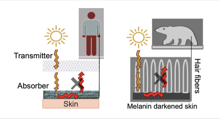
A new double-layered fabric inspired by the black skin and white fur of polar bears uses heat radiated from the Sun and indoor lighting to trap and maintain warmth. The fabric could be used to make “personal climate” textiles that weigh 30% less than cotton for a given area and keep wearers warm at temperatures 10 °C colder.

Although fabric technologies have advanced significantly in recent years, a truly thermoregulating textile is still lacking. The new material, created by researchers at the University of Massachusetts Amherst in the US, partly addresses this gap by pairing an outer layer that transmits visible light with a base layer that absorbs it while reflecting light at infrared frequencies.

“The outer layer receives light from the Sun and then transmits this light down to the base layer,” explains team leader Trisha L Andrew, a chemist and wearable electronics expert at Amherst. “This is much like how a polar bear’s fur, which is essentially a natural fibre optic, conducts sunlight down to the bears’ skin. The skin absorbs the light, heating the bear.”

On-body greenhouse effect

Polar bears evolved their combination of dark skin and white hair to keep warm in their icy habitat by selectively reflecting, absorbing or transmitting radiation across the visible and infrared parts of the electromagnetic spectrum. Though the precise details of the mechanism are not well studied, scientists believe that low optical density (light-coloured) insulating features such as white fur help polar bears achieve an on-body version of the greenhouse effect.

Somewhat counterintuitively, this white fur transmits much more radiation to the bear’s skin than darker hair would. Indeed, the “solar utilization factor” – a ratio of heat utilized to total solar heat gain – for polar bears ranges from 10% to 50%. This high utilization is further enhanced by the polar bear’s skin, which is rich in melanin – a dense biopolymer made up of conjugated units that have a high refractive index and absorb light at a broad range of wavelengths.

Similar thermoregulating surfaces are also present in other animals, including species of moths, butterflies and birds that have adapted to live in cold but sunny climates. These surfaces selectively absorb light in the visible and near-infrared part of the spectrum (where photothermal heating occurs), while reflecting it over the infrared range, where objects spontaneously radiate heat according to Planck’s law.

Some artificial materials also regulate heat in this way, and researchers have previously made textile coatings from optical materials such as MXene, carbon nanotubes and silver nanowires. However, the resulting fabrics did not contain a visible-transmitting outer layer to prevent heat from being lost to the wearer’s surroundings.

An alternative to inorganic or carbon nanomaterials

In designing their new base layer, Andrew and colleagues eschewed inorganic or carbon nanomaterials in favour of nylon coated with a dark polymer called poly(3,4-ethylenedioxythiophene), or PEDOT. This polymer is particularly well-suited to making textiles and has a high optical density while remaining lightweight and flexible.

To test the new thermal textile, the Amherst team used skin and solar simulators. These measurements showed that when the textile is exposed to a moderate light intensity of 130 W/m2, it keeps its wearer just as warm as cotton fabric would – and at temperatures that are 10 °C colder – while weighing 30% less.

“Our polar bear fabric could be very useful for managing space heating, which consumes huge amounts of energy, in a more energy-efficient manner, by heating people indoors using ambient lighting instead of room heating,” Andrew tells Physics World.

“By focusing energy resources on the ‘personal climate’ around the body, this approach could be far more sustainable than the status quo,” adds study lead author Wesley Viola.

The Amherst team is now exploring how to achieve on-body radiative cooling, which will be vital as climate change pushes temperatures higher and heat waves become more common even in temperate regions.

The new thermal textile is described in ACS Applied Materials & Interfaces.

Copyright © 2026 by IOP Publishing Ltd and individual contributors