The events, which include lectures, demonstrations and careers talks, will take place at Imperial College London, the National Physical Laboratory in Teddington and the Institute of Physics in London.
To apply for the programme, you will need to be a student of Black or mixed-Black heritage, in Year 11 or 12 and based in London.
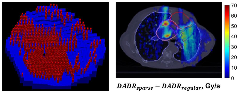
Optimizing protons for FLASH Left: sparse ridge filter design. Right: the difference in dose-averaged dose rate (DADR) between a sparse ridge filter and regular ridge filter. (Courtesy: Int. J. Radiat. Oncol. Biol. Phys. 10.1016/j.ijrobp.2023.01.048)
Stereotactic body radiation therapy (SBRT) is a precision cancer treatment that delivers fewer, higher-dose radiation fractions than traditional radiotherapy. SBRT can provide excellent local tumour control, but for some tumour locations it risks exposing nearby organs-at-risk (OARs) to unacceptable irradiation levels. Proton-based SBRT provides better OAR sparing, but still requires some treatment margins that may limit its clinical applicability.
FLASH radiotherapy, in which radiation is delivered at ultrahigh dose rates, could enable further sparing of OARs. To investigate its potential, a research team headed up at Emory University is developing a framework for optimizing proton therapy delivery to meet the demands of FLASH radiotherapy.
Most modern proton therapy systems can achieve FLASH dose rates using a high-energy transmission beam that passes through the patient, depositing dose throughout its path. This approach, however, eliminates the major advantage of proton therapy: its ability to deliver dose in a spread-out Bragg peak. To improve conformality at FLASH dose rates, Ruirui Liu and colleagues propose that patient-specific ridge filters could provide a similar dose distribution to that of conventional intensity-modulated proton therapy (IMPT).
For FLASH treatments, the dose, dose-averaged dose rate (DADR) and dose-averaged linear energy transfer (LETd) all influence the biological response. Thus the researchers developed an integrated physical optimization (IPO) framework that simultaneously optimizes these three parameters to maximize OAR sparing in a patient’s treatment plan. The framework, described in the International Journal of Radiation Oncology, Biology, Physics, uses the IPO-IMPT objective function to provide multiple solutions for the design of patient-specific ridge filters and proton spot maps.
The ridge filters, which are used in combination with a range compensator, comprise an array of ziggurat-shaped pins that spread the Bragg peak from a 250-MeV beam to cover a beam-specific planning target volume. The team developed inverse planning software to define the pin locations for a patient-specific filter, and used Geant4-based Monte Carlo simulations to provide dose and LET influence matrices.
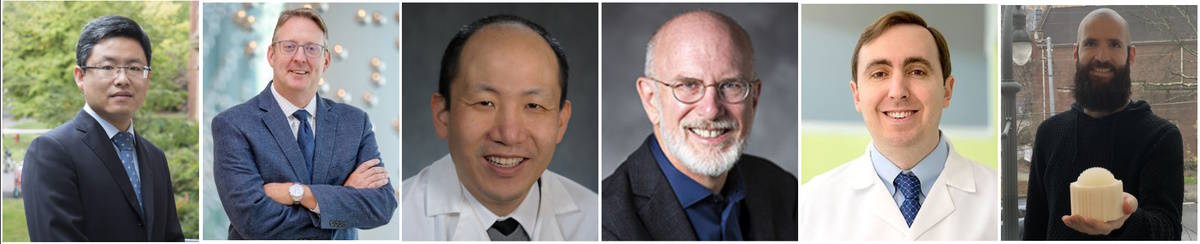
Research team From left to right: Ruirui Liu, Jeffrey Bradley, Liyong Lin, William Dynan, Charles Simone II and Nathan Harrison, holding the ridge filter. (Courtesy: Nathan Harrison)
Patient plans
To demonstrate the IPO-IMPT framework, the researchers developed treatment plans for three patients with lung cancer. They prescribed a dose of 50 Gy (five 10 Gy fractions) to the clinical target volume, with a maximum hotspot dose of 62.5 Gy. Depending on which parameter is prioritized, the plans aim to increase FLASH coverage and/or reduce LETd, while maintaining target dose.
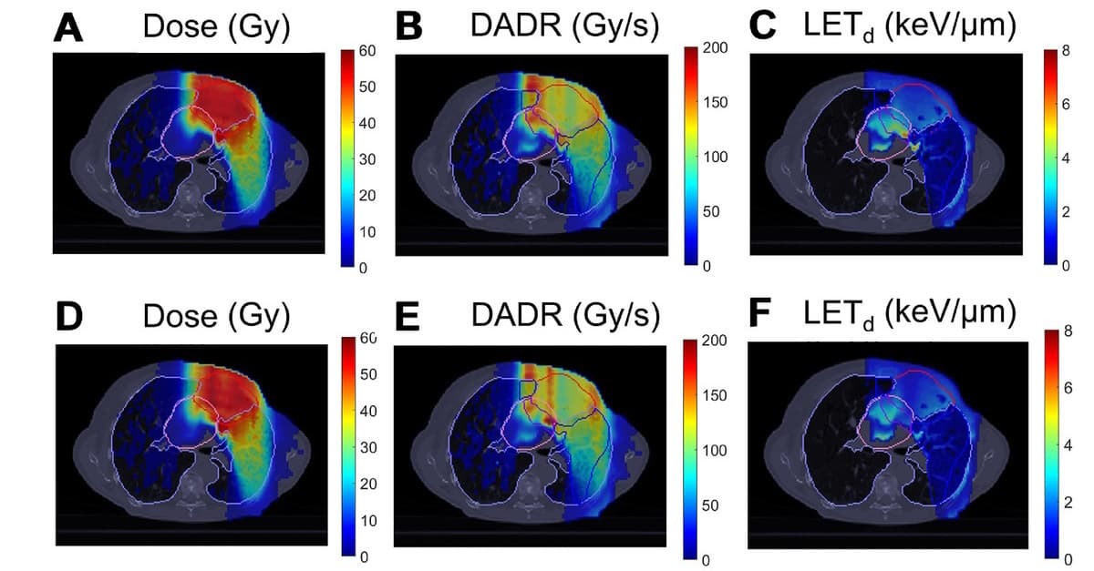
For patient 1, who had a central lung tumour close to the heart, the OARs were the heart and lung. For this case, the researchers generated a single-beam IPO-IMPT plan with the aim of reducing LETd to the heart while maintaining target coverage. The IPO-IMPT plan met this goal, exhibiting similar target coverage to a conventional IMPT plan but markedly reducing LETd to the heart.
Plan analysis Treatment plans for patient 1 using a regular ridge filter. (A-C) Dose, dose-averaged dose rate (DADR) and dose-averaged linear energy transfer (LETd) distributions for IMPT. (D-F) Dose, DADR, and LETd distributions for IPO-IMPT. (Courtesy: Int. J. Radiat. Oncol. Biol. Phys. 10.1016/j.ijrobp.2023.01.048)
Patient 2 had a metastatic tumour in the right lower lobe and patient 3 had a tumour in the subcarinal lymph node. In these cases, the oesophagus was also an OAR and the key goal was oesophageal sparing. For both IPO-IMPT and IMPT, almost 100% of the oesophagus evaluation volume met the 40 Gy/s FLASH threshold, For patient 2, IPO-IMPT slightly decreased LETd for the heart and oesophagus and increased FLASH coverage for the heart.
Sparse pin design
Regular ridge filters designed using the IPO-IMPT framework selectively spared OARs by reducing LET and increasing FLASH coverage. However, sparse ridge filters, from which some pins are omitted, offer potential to further increase OAR sparing. Removing filter pins at specific locations delivers a higher proton flux, while the remaining pins still provide adequate target coverage.
For patient 1, the researchers generated an IPO-IMPT plan with sparse ridge filters and multiple beams. Comparison with an IMPT plan using regular ridge filters showed that, for both, tumour coverage was maintained and hotspots were well controlled. The sparse ridge filters, however, increased the OAR volume receiving a FLASH dose rate by 31% and 50%, for heart and lung evaluation volumes, respectively.
The sparse ridge filters provide flexibility to realize the full potential of the IPO-IMPT framework. For example, the pin removal levels can be tailored to individual patient cases. A 50% pin removal threshold provided reasonable results for patient 1’s large tumour, while a 30% threshold was a good starting point for the smaller targets of patients 2 and 3, whose sparse ridge filter-based plans increased DADR in the oesophagus while maintaining tumour coverage.
Finally, to verify that a ridge filter assembly (filter pins and a compensator) could deliver the predicted dose, the researchers 3D printed a patient-specific ridge filter. They delivered a treatment plan designed to provide a uniform target dose and performed dose measurements with an ionization chamber array. The total gamma passing rate was 92.9% for absolute doses, which exceeds the standard patient passing criteria of 90% and demonstrates that the assembly can deliver a clinically acceptable dose distribution.
“This proof-of-concept study demonstrates the feasibility of using an IPO-IMPT framework to accomplish FLASH stereotactic body proton therapy, accounting for dose, DADR and LETd simultaneously,” the researchers conclude. “This novel method will facilitate delivery of conformal proton fields at FLASH rates for preclinical and clinical studies.”
Senior author Liyong Lin tells Physics World that the team hopes to further develop its software for such applications. “Emory’s Office of Technology Transfer encouraged us to form a startup company, Radiotherapy Biological Optimization (RBO) Solutions,” Lin explains. “RBO is accepted by the National Institutes of Health’s Applicant Assistance Program to submit a small business technology transfer R41 grant to the National Cancer Institute by April 5. IBA, the biggest particle therapy vendor, and IBA’s dosimetry division will endorse RBO’s R41 grant proposal.”
Update 29/03/2023: Astronomers have reported the results of their observatios on GRB 221009A in a special issue of Astrophysical Journal Letters. The article below was first published on 14/10/2022.
Several orbiting space telescopes scanning the skies for powerful cosmic explosions have spotted one of the brightest gamma-ray bursts ever detected. Initial evidence suggests that the blast of high-energy radiation occurred when an extremely massive star collapsed – a process that results in an immense flood of gamma-rays and X-rays. Astronomers have been racing to follow-up the discovery, with one researcher suggesting it will become the “best studied gamma-ray burst in history”.
The first reports of the explosion, catalogued as GRB 221009A, came from the Neil Gehrels Swift Observatory and the Fermi Gamma-ray Space Telescope, which both monitor the universe at gamma-ray and X-ray wavelengths. Their systems noticed a bright source appear in the constellation Sagitta on 9 October. The blast was also picked up by the European Space Agency’s Solar Orbiter mission and since then numerous other observatories – including those looking at visible wavelengths – have scrutinized the fading fireball from the event, which is known as the “afterglow”.
One of the most striking aspects about GRB 221009A is its proximity. The blast appears to have happened in a galaxy about two billion light years away, which is considerably closer than an “average” gamma-ray burst event that may lie some 10 billion light years away. Leicester University astronomer Kim Page, who works on NASA’s Swift mission, says that such closeness had “a big part to play” in why this burst appeared so bright.
The bright and nearby nature of GRB 221009A should give scientists plenty to study. “We have a lot of photons across the electromagnetic spectrum, and this will allow us to slice up the data more finely,” adds Page. Researchers will also be hoping to examine how the chemical signature, or spectrum, of the explosion evolves – information that can reveal clues about the composition of the phenomenon.
A ‘unique’ measurement
While initial analyses are still ongoing, astronomers are already marvelling at some of the early observations. X-ray imagery from the Swift observatory shows prominent, glowing rings around the location of GRB 221009A. These features are not physically part of the blast but “light echoes” that are caused when X-ray radiation streaming from the event scatters towards Earth off microscopic grains suspended within dust clouds inside our own galaxy.
GRB 221009A is going to be the best studied gamma-ray burst in history
Andrea Tiengo
“This is by far the best set of rings seen around a gamma-ray burst, thanks partly to its brightness in X-rays and its closeness to the Galactic plane,” explains Leicester astronomer Andrew Beardmore, who works on the Swift mission.
Beardmore says analysis of the rings will allow scientists to investigate the nature of the interstellar dust grains and even probe the locations of the Milky Way dust clouds where they reside. “Because [the rings] are so bright, the distance to the dust-layers responsible for [them] will likely be known with great precision. This makes it quite a unique measurement,” he adds.
Indeed, that measurement is something that Andrea Tiengo from the Scuola Universitaria Superiore IUSS di Pavia, in Italy, and colleagues have already been working on. “We have compared our measurements with the distances derived from other methods and we confirm that they are compatible,” he says. “But we detect more clouds up to a larger distance and determine their distance with higher accuracy.”
The dust responsible for the rings has also scattered a particular kind of X-ray light from the early stages of the blast. These so-called “soft” X-rays – with energies of between 0.3 and 10 keV – aren’t usually emitted towards us during these events, but they can be studied for details about the star that formed the gamma-ray burst.
“Since GRB 221009A is going to be the best studied gamma-ray burst in history, this is a fundamental piece of information that would otherwise be missing and it will certainly help us to better understand the physics of the most powerful explosions in the universe,” says Tiengo.
Extended lifetime Quantum information is protected by encoding it in a more complicated system, such as the “GKP state”. (Courtesy: Volodymyr Sivak, Yale University)
A team of researchers at Yale University has been able to observe and correct errors in real time as they occur in a quantum system, preserving the information for over twice as long as it could otherwise be stored.
Quantum computing uses certain physical systems, such as superconducting circuits, atoms, ions or photons, to perform calculations that would be too difficult for classical computers. However, quantum states are delicate and susceptible to noise, making them difficult to maintain. Researchers have proposed various methods that correct these errors as they occur; however, all come with considerable hardware challenges.
Quantum information is usually stored as qubits, using quantum systems that can be in a “superposition” of two different states at the same time, such as a photon with two different polarization states. All error-correction schemes need to make use of extra information in the system, as this information is employed to notify the observer that errors have occurred and give details that are used to correct them. These schemes can make use of extra states in the same quantum system hosting the qubit, such as additional energy levels, or they can combine several qubits together.
Optimizing the experiment
The team, led by Michel Devoret, used the former method, employing a qubit encoding known as a GKP (Gottseman-Kitaev-Preskill) code. This was implemented using light confined in an aluminium cavity, connected to a sapphire chip with a superconducting qubit (known as a “transmon”). The cavity was used as the main system to store the quantum information, which was controlled by the transmon.
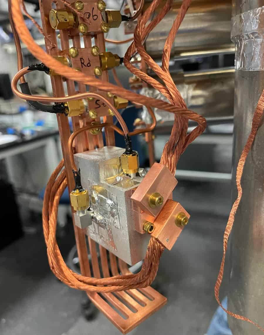
Keep it cool This image shows copper braids that run between the dilution refrigerator, used to maintain the system at extremely low temperatures, and the clamp that holds the transmon in place. (Courtesy: Volodymyr Sivak, Yale University)
In order to accurately assess the improvement gained by using error correction, the lifetime of the qubit had to be compared with the uncorrected lifetime of other potential qubits in the system. This could be either using the transmon itself, or using the cavity as a simple two-level system (instead of preparing the full GKP state), which had the longest lifetime of 800 µs. The extended lifetime of the GKP qubit measured after error correction was 1.82 ms, an improvement of 2.27 times.
Each cycle of measuring and correcting errors takes 10 µs, requiring state-of-the-art hardware. To optimize the experimental parameters, the team employed a machine-learning system to choose, for example, the average number of photons used when implementing operations.
A lower number of photons was chosen, which increases the time for these procedures, but reduces the probability of harder-to-correct errors. Optimization like this could be a valuable tool to deal with the complexity of future experiments when choosing parameters.
As lead author Volodymyr Sivak, now a research scientist at Google, explains, “other experiments normally track the error, but do not intervene to correct it”. As an experimental demonstration that it is truly possible to extend the lifetime of a quantum state using error correction, this is a major milestone for quantum computing, according to Sivak, “to show that there is no fundamental obstacle to doing quantum error correction, that it is actually possible in the real world and not only on paper”.
Other error-correction schemes require many qubits, which need to pass a certain quality threshold before they can be combined to reduce the overall error. Future work could involve using a GKP qubit such as the one introduced in this work as a “base qubit” for these techniques, although as Sivak notes, this “will take some time”.
For the first time, pure water has been used to detect low-energy antineutrinos produced by nuclear reactors. The work was done by the international SNO+ collaboration and could lead to safe and affordable new ways to monitor nuclear reactors from a distance.
Situated 2 km underground near an active mine in Sudbury, Canada, the SNO+ detector is the successor to the earlier Sudbury Neutrino Observatory (SNO). In 2015, SNO’s director Art McDonald shared the Nobel Prize for Physics for the experiment’s discovery of neutrino oscillation – which suggests that neutrinos have tiny masses.
Neutrinos are difficult to detect because they rarely interact with matter. This is why neutrino detectors tend to be very large and are located underground – where background radiation is lower.
At the heart of SNO was a large sphere of ultra-pure heavy water in which energetic neutrinos from the Sun would very occasionally interact with the water. This produces a flash of radiation that can be detected.
Careful measurements
SNO is currently being upgraded as SNO+, and as part of the process ultra-pure normal water was temporarily used as the detection medium. This was replaced by a liquid scintillator in 2018, but not before the team was able to made a series of careful measurements. And these threw up a surprising result.
“We found our detector was performing beautifully, and that it might be possible to detect antineutrinos from distant nuclear reactors using pure water,” explains Mark Chen. He is the SNO+ director and is based at Queen’s University in Kingston, Canada. “Reactor antineutrinos have been detected using liquid scintillators in heavy water in the past, but using just pure water to detect them, especially from distant reactors, would be a first.”
It had been difficult to detect reactor antineutrinos in pure water because the particles have lower energies than solar neutrinos. This means that the detection signals are much fainter – and therefore are easily overwhelmed by background noise.
Lower background
As part of SNO+’s upgrades, the detector was fitted with a nitrogen cover gas system, which significantly lowered these background rates. This allowed the SNO+ collaboration to explore an alternative approach to detecting reactor antineutrinos.
The detection process involves a neutrino interacting with a proton, resulting in the creation of a positron and a neutron. The positron creates an immediate signal whereas the neutron can be absorbed sometime later by a hydrogen nucleus to create a delayed signal.
“What enabled SNO+ to accomplish this detection are very low backgrounds and excellent light collection, enabling a low energy detection threshold with good efficiency,” Chen explains. “It’s the latter – a consequence of the first two features – that enabled the observation of antineutrinos interacting in pure water.”
“Dozen or so event”
“As a result, we were able to identify a dozen or so events that could be attributed to interactions from antineutrinos in pure water,” says Chen. “It’s an interesting result because the reactors that produced those antineutrinos were hundreds of kilometres away.” The statistical significance of the antineutrino detection was 3.5σ, which is below the threshold of a discovery in particle physics (which is 5σ).
The result could have implications for the development of techniques used to monitor nuclear reactors. Recent proposals have suggested that antineutrino detection thresholds could be lowered by doping pure water with elements like chlorine or gadolinium – but now, the results from SNO+ show that these costly, potentially dangerous materials may not be necessary to achieve the same quality of results.
Although SNO+ can no longer make this type of measurement, the team hopes that other groups could soon develop new ways to monitor nuclear reactors using safe, inexpensive, and easily attainable materials, at distances that will no disrupt reactor operation.
When pioneering musicians such as Kraftwerk and Brian Eno began experimenting with synthesizers and digital samplers in the 1970s, it was considered avant-garde and confined to niche audiences. It didn’t take long, however, for electronic music to explode in popularity, and today computer-produced music is ubiquitous among many genres and styles. This episode of the Physics World Stories podcast looks at a new trend in its nascent stages – music generated by quantum computers.
Later in the episode, podcast host Andrew Glester is joined by Maria Mannone, a theoretical physicist working on quantum information at the University of Palermo in Italy, who is also a composer. Mannone discusses some of her experiments that incorporate scientific concepts into sound, and you can hear some of the music that emerges.
For much more quantum-inspired content, make sure to visit this website again on 14 April for World Quantum Day. During that week, the Physics World Weekly podcast will have a quantum theme and we will share a selection of quantum-related feature articles, interviews and analysis pieces. There will also be a chance to access quantum content and discounted quantum ebooks, shared by IOP Publishing – which publishes Physics World.
This episode is sponsored by Pfeiffer Vacuum. The company provides all types of vacuum equipment, including hybrid and magnetically-levitated turbopumps, leak detectors and analysis equipment, as well as vacuum chambers and systems. You can find about Pfeiffer Vacuum’s impact in space research in this video, and explore all its products on the Pfeiffer Vacuum website.
Researchers who are not white face “significant” inequalities when publishing their work, including longer publication delays than white scientists and fewer overall citations. That is according to a new analysis in the Proceedings of the National Academy of Sciences, which examined a million scientific papers published between 2001 and 2020. It also finds that Black researchers in the US are the most under-represented on journal editorial boards.
The study was carried out by Fengyuan Liu – a computer scientist at New York University Abu Dhabi – and colleagues Talao Rahwan and Bedoor AlShebli. The papers in the study were sourced from more than 500 journals from six different publishers: Frontiers Media, Hindawi, the Institute of Electrical and Electronics Engineers, the Multidisciplinary Digital Publishing Institute, Public Library of Science and the National Academy of Sciences.
For each paper, the researchers identified the authors and the journal editors who handled the paper during the submission process, the latter being made up of nearly 65,000 people. Bibliometric information on both groups was accessed via the Microsoft Academic Graph dataset with a tool known as NamePrism being used to classify the scientists into one of six racial groups based on their names.
The study showed that scientists from the bulk of nations in Africa, Asia and South America — where most people are ethnically non-white — are under-represented on editorial boards relative to their representation among paper authors. Articles by researchers from these regions also generally have longer gaps between submission and acceptance compared to articles other published in the same journals during the same year.
‘A grim picture’
The researchers also found that papers by non-white scientists receive fewer citations than would be expected based on comparable articles by their white peers. The lower rate of citation will make those scientists less visible in the community find it harder to secure grants and awards.
When Liu and colleagues examined US-based scientists, they found Black researchers are the most under-represented on editorial boards and suffer from the longest delays between paper submission and publication. Black and Hispanic scientists in the US, meanwhile, receive far fewer citations than their white peers doing similar research.
In physics, the team found that around 80% of countries from Africa, Asia and South America are under-represented on editorial boards — a figure that is typical across the sciences. In contrast, European, North American and Oceanian countries are almost equally divided between being over- and under-represented on editorial boards.
The researchers say their findings “paint a grim picture in which non-white scientists suffer from inequalities that may hinder their academic careers”. To address these disparities, the authors call on journal publishers to review how they select board members, the time it takes to review submissions, and how they promote published manuscripts.
“The responsibility to take action falls not only on the shoulders of the publishers, but also on the scientific community as a whole to create an ecosystem without geographic and racial disparities,” the authors write.
As global demand for electrification grows, so does the need for reliable, affordable batteries. Battery technology will be pushed to meet these demands over the next 10–15 years, and developers with cutting-edge techniques will have a significant advantage.
Specifically, battery developers are tasked with improving five key qualities in their products:
Double the energy density
Quadruple the power density
Make batteries safer
Double their lifespan, and
Reduce cost of electrode manufacturing and halve the cost for each cell.
Achieving these breakthroughs in battery technology will require thorough material characterization and development. Understanding and optimizing battery materials is possible through leveraging multiple powerful material-analysis techniques.
This webinar will cover the essential material characterization techniques that today’s labs need to adopt to develop the batteries of tomorrow.
Hang Kuen Lau, TA Instruments – Waters LLC
Hang Kuen Lau, PhD, is a segment field marketing manager focused on the battery market. Hang joined TA Instruments in 2018 as an applications engineer supporting the thermal product lines and has worked as a scientific lead in new market development for batteries. She received her PhD in materials science and engineering from the University of Delaware and earned her BS/MS from Drexel University.
Alexandra Stavropoulou, Oxford Instruments NanoAnalysis
Alexandra Stavropoulou is a geologist by training. She studied in the National and Kapodistrian University of Athens and in Trinity College Dublin. The study of minerals and nature has always intrigued her, initially emphasising on metals (gold and manganese occurrence), later expanding to rock texture analysis. Microanalytical approaches, especially scanning electron microscopy (SEM) coupled with energy dispersive spectroscopy (EDS) and electron backscatter diffraction (EBSD) opened up new doors for Alexandra in the observation and quantification of minerals and rocks properties. She joined Oxford Instruments in 2020 as a segment marketing scientist, focusing on battery materials characterization.
Some people know from a young age that they want to be a scientist and that – with enough ability and effort – they can reach that goal. Gertrude Scharff (Scharff-Goldhaber after she married) felt that early calling. And while she had the ability to fulfil it, her path to scientific success had more than its share of personal hardships and professional obstacles.
Born to a German-Jewish family on 14 July 1911, she lived through the First World War, the post-war upheavals in Germany and the rise of Hitler. After earning her PhD in physics from the University of Munich, she sought entry into a profession dominated by men. When she fled Nazism, she faced difficulties as an immigrant to the UK. And when she tried to build a new life in the US with her physicist husband, she still struggled to find scientific employment, as the rigid rules of nepotism thwarted her career.
Yet she endured, and established herself as a highly respected nuclear physicist, one of the few pioneering women in that area. Her research advanced the understanding of nuclear fission and contributed to the theory of nuclear structure. Her work was recognized in 1972 when she became only the third female physicist elected to the National Academy of Sciences. She is also well remembered as an advocate for women in science, for encouraging young scientists and for championing science education.
Ominous times, outstanding student
Known as Trude to her friends and family, Scharff’s early years in Germany were turbulent ones, encompassing the First World War, political unrest and economically ruinous hyperinflation after the country’s defeat in 1918. At the age of eight she saw Communist revolutionaries being slaughtered by the military in the streets of Munich, where her family lived. Later she would recall having to eat bread bulked out with sawdust. The turmoil continued, with ominous forebodings for German Jews, as Hitler rose to power in 1933.
Tumultuous time Nelly Scharff posing with her two young children Gertrude (left) and Liselotte (right). Painted by Friedrich August von Kaulbach in Munich. Nelly Scharff and her husband Otto died during deportation by the Nazis in 1941. Their daughters had already left Germany safely. (Courtesy: Leo Baeck Institute)
Amid all this, Scharff obtained a worthy education. According to a memoir by her son Michael, she attended an elite high school for girls. An outstanding student, she developed an interest in physics. Her father had hoped she would study law to prepare for managing the family business, but she was more keen to “understand what the world is made of”, as she later put it.
But outside events utterly changed her plans and her life. As Nazism spread, Scharff found herself ostracized by colleagues and German Jews began fleeing the country. She was, however, well along in her research. As she told an interviewer in 1990: “I should have left earlier. But since I had started my thesis, I felt I should finish.”
Finish she did, in 1935, but she cut it very close. That was the year the Nuremberg Laws were enacted, defining first Jews and later Romani and Black Germans as “inferior races” and “enemies of the state”. They were effectively barred from German society, and faced harsh penalties for violating the laws. Antisemitic violence grew and Scharff’s parents later perished in the Holocaust.
Aware that it was most certainly time to escape from Germany, Scharff wrote to 35 refugee scientists seeking a position elsewhere. Almost all told her not to come because there was already a glut of refugee scientists – except for Maurice Goldhaber, a young Austrian-Jewish physicist she had met in Germany. Working on a PhD at the University of Cambridge under Ernest Rutherford, he thought there could be opportunities in England. Moving to London, Scharff eked out a living for six months by selling a prized possession that was part of her wedding trousseau – a Leica camera, well known for its fine optics – and translating articles from German into English. Then she worked at Imperial College London under George Thomson, studying electron diffraction (in 1937 he shared the Nobel Prize with Clinton Davisson for discovering the effect in crystals), but never found an independent research position.
In 1939 her prospects improved. Scharff married Goldhaber, becoming Scharff-Goldhaber, and the couple moved to the US. Goldhaber had a faculty position at the University of Illinois-Urbana, but Scharff-Goldhaber could not become a full-fledged academic scientist, because anti-nepotism laws in Illinois did not allow the university to hire her. She could do research only as an unpaid assistant in her husband’s lab. This moved her from condensed-matter physics into his field of nuclear physics. Scharff-Goldhaber’s papers in the 1940s produced under these circumstances show that she handled the transition brilliantly – but she never reached full faculty status at Illinois.
A new lab on Long Island
Only in 1950 did Scharff-Goldhaber and her husband together find a true research home, at the new Brookhaven National Laboratory (BNL), which had been founded three years earlier. Today a US Department of Energy facility, the lab’s original mandate was to seek peaceful uses of atomic energy. Its scientific efforts have since diversified but nuclear and high-energy physics remain part of its research activities.
Her appointment made Scharff-Goldhaber the first female physicist at BNL, and 15 years after earning her degree, she was finally being paid as a professional researcher. Even so, she operated in an atmosphere that her son Michael describes as only “grudgingly accepting”. Goldhaber was hired as a “senior scientist” and ran his own research group, but Scharff-Goldhaber was ranked simply as a scientist within his group. (Goldhaber would eventually rise to director of the lab 1961–1973, and Scharff-Goldhaber to senior scientist.)
As the only woman with professional scientific status at BNL, Scharff-Goldhaber had no female scientific peers. Most women associated with the lab were the non-working wives of male scientists, who in the 1950s filled traditional roles. With two children, Michael and Alfred, Scharff-Goldhaber had similar responsibilities; but at social events she was more likely to talk physics with the men than discuss childcare with the women. Within this male milieu she formed good relationships with her colleagues, and with the support staff who produced the isotopes she needed for her research at the BNL reactor or Van de Graaff accelerator.
Fission, and a fundamental experiment
Except for the period in the 1930s when she was still trying to become an independent scientist, Scharff-Goldhaber maintained a brisk pace of research and publication, while meeting family obligations. In 1936 she published “The effect of stress on the magnetization above the Curie point” from her thesis. Her next set of papers began four years later, once she switched to nuclear physics in 1940 at Illinois, and she wrote over a dozen more until she was fully settled at BNL. Over the next 30 years, she published some 60 more papers, mostly in the Physical Review, and contributions to conference proceedings.
Several of the papers arising from her work at Illinois in the 1940s are especially notable, including one that concerned spontaneous nuclear fission. In 1938 Lise Meitner and Otto Frisch had found that a uranium nucleus bombarded with neutrons could split in two and release much energy. If neutron-induced fission could be made self-sustaining, it could produce an enormously destructive weapon. With war looming, European and American physicists investigated self-sustaining fission hoping that the Nazis would not find the answer first.
Chain reaction In 1942 Gertrude Scharff-Goldhaber demonstrated that uranium undergoing spontaneous fission released neutrons along with energy. These neutrons could activate more nuclei and more energy, with the phenomenon used by Manhattan Project scientists to build an atomic bomb. (Courtesy: iStock/Andreus)
In 1942 Scharff-Goldhaber directly showed, apparently for the first time, that uranium undergoing spontaneous fission released neutrons along with energy. These neutrons could activate more nuclei and more energy – a cascading chain reaction that could become a nuclear explosion. Data like these were crucial to achieving the world’s first self-sustaining controlled nuclear reaction in 1942, as the atomic bomb was being built by the Manhattan Project. The Scharff-Goldhabers were not yet US citizens and so were not part of the project, but her result was secretly circulated to relevant scientists and was published after the war (Phys. Rev.70 229).
In a separate paper published in 1948 (Phys. Rev. 73 1472), the Scharff-Goldhabers together answered a fundamental question: are beta rays exactly the same as electrons? Discovered in 1897 in cathode rays by J J Thomson, electrons were the first known elementary particles. A few years later in 1899, Rutherford was studying the new phenomenon of radioactivity, and found an unknown emission he called beta rays. These turned out to be charged particles with the same charge to mass ratio e/m as electrons and were identified as such. But the question remained: could beta rays and electrons differ in some other property such as spin?
The Scharff-Goldhabers cleverly tested this hypothesis by using the Pauli exclusion principle, which, they wrote, “would not hold for a pair of particles if they differed in any property whatsoever”. In their experiment, they irradiated a lead sample with beta rays. If these were not identical to electrons, they would not obey the Pauli principle. Then they would be captured by lead atoms, enter bound orbits already filled with electrons, and transition to the lowest orbit, causing X-rays to be emitted. If beta rays and electrons were identical, the former would be barred from entering atomic orbits and producing X-rays. The experiment detected no X-rays at the expected energies, confirming that beta rays are electrons emitted from radioactive nuclei.
Excited nuclei and “magic” numbers
Starting in the early 1950s at BNL, Scharff-Goldhaber began what would be her career-long project: to form a systematic picture of the properties of excited nuclei across the periodic table. Her plan to work in “low-energy” nuclear physics diverged from her husband’s growing interest in “high-energy” physics, where huge new particle accelerators probed fundamental particles. According to their son Michael, Scharff-Goldhaber’s separate path deprived his father of her great abilities as an experimentalist. But he adds that “the split did not prevent the family dinner table conversation from focusing on nuclear physics, just as it had before, largely to the bafflement of the children”. (Later he and Alfred each earned a doctorate in theoretical particle physics.)
At the time, the behaviour of the excited nucleus was just beginning to be grasped. This dense soup of protons and neutrons could be viewed as a collection of particles bound together by nuclear forces, forming a medium with an energy that is expressed in rotation or vibration of the entire body. In the so-called “shell model”, however, the nucleus was seen as a quantum system where nucleons occupy energy levels, analogous to the discrete levels or “shells” occupied by electrons in an atom. Each approach had successes. Treating the nucleus as a liquid led to an understanding of how it could deform and undergo fission. The shell model predicted that nuclei with specific, or “magic”, numbers of protons or neutrons (2, 8, 20, 28…) would be exceptionally stable, again analogous to filled electronic shells in atoms.
Family business Alfred Goldhaber and his mother Gertrude Scharff-Goldhaber co-wrote two papers in nuclear physics. (Courtesy: AIP Emilio Segrè Visual Archives, Physics Today Collection)
It wasn’t clear, however, whether experiment really supported the shell model, or where each approach could best be applied. Scharff-Goldhaber’s extensive research on different nuclei helped to resolve these issues. Her work was significant in developing the theory that finally connected the two approaches, which led to Aage Niels Bohr, Ben Mottelson and Leo Rainwater sharing the 1975 Nobel Prize for Physics.
In the 1950s Scharff-Goldhaber measured the energy of excited nuclei versus neutron number and showed that shell structure influenced the energy, which peaked at the magic numbers. She also noted an anomalous change in energy levels with an increase in the number of neutrons, which she related to a change in shape of the nucleus. Later she developed her own “variable moment of inertia” (VMI) model, which used the shape of nuclei to provide further insight into their energies across the periodic table.
Besides her contributions to nuclear theory, Scharff-Goldhaber’s research in this era had unusual features. She wrote two papers about the VMI model together with her son Alfred – as far as is known, the only mother–son research papers in physics (Phys. Rev. Lett.24, 1349 ; Phys. Rev. C17, 1171).
She also enhanced her data analysis by extending the standard nuclide chart, where each nucleus is placed in a two-dimensional plot of number of protons versus number of neutrons. Scharff-Goldhaber glued vertical rods of length proportional to the lowest excitation energy for each nuclear species to the appropriate position on the chart. Long before the routine use of 3D computer visualizations, this was a tremendous aid in spotting important features such as the energy change between N = 88 and N = 90.
Innovative teacher Gertrude Scharff-Goldhaber in her office at Brookhaven National Laboratory. Mounted on the wall near the top of the photo is one of her 3D nuclide charts. (Courtesy: Gertrude S Goldhaber Collection, Leo Baeck Institute, AR 25820 A)
Along with her research, Scharff-Goldhaber found ways to help women in science, and to contribute to science education and the scientific community. Among many professional involvements, she served on American Physical Society (APS) committees devoted to the status of women in physics and to pre-college physics education. She was known too for reaching out to early career scientists – both men and women. One was Rosalyn Yalow, Goldhaber’s PhD student at Illinois, who shared the 1977 Nobel Prize in Physiology or Medicine for inventing the radioimmunoassay technique. Yalow has credited both her adviser and Scharff-Goldhaber “for support and encouragement”. Scharff-Goldhaber also broadened the intellectual atmosphere at BNL by founding the Brookhaven Lecture Series, featuring eminent speakers such as Richard Feynman.
Retired, but still researching
Scharff-Goldhaber had started at BNL relatively late and was ready to continue her research for a long time, but the stringent retirement laws of the era officially ended her employment in 1977, aged 66. According to her son Michael, the retirement was forced in a way that he calls “subtly sexist”. Nevertheless, working without pay, she collaborated with other scientists and co-authored research papers until 1988. When failing health restricted her activities, however, she appreciated and sought satisfaction in what she could still do, until she died at the age of 86 in 1998.
In 1990 a journalist interviewing Scharff-Goldhaber noted her “soft but insistent determination” – likely the very character traits that enabled her to overcome barriers to a research career. In 2016, looking back on his mother’s life, Michael described her as “a person of unique wilfulness and even stubbornness, traits that she certainly needed…to pursue a successful career in a world that was often set against her”.
Perhaps Scharff-Goldhaber would agree with these assessments, but there’s another one that I believe applies. In 1972, reviewing a book about nuclear energy by Isaac Asimov, Scharff-Goldhaber wrote that progress in science, among other qualities, is “based on the burning desire to get to the bottom of things”. Writing those words, did she reflect that her own life perfectly exemplifies that ethos?
Metrologists enable radiation therapy. These experts in measurement science develop measurement standards and calibrate detectors used in radiotherapy clinics, and when new treatment modalities such as FLASH proton radiotherapy are introduced, they establish accurate dosimetry for those systems.
Dosimetry for FLASH beams, which deliver radiation quickly using ultrahigh dose rates, is complicated. Ionization chambers, detectors ubiquitous in the radiation therapy community, exhibit ion recombination effects under FLASH beams. As a result, ionization chambers used in FLASH applications require large corrections with large uncertainties that reduce scientists’ confidence in knowing how much radiation dose has been delivered.
“At NPL, we’re trying to disseminate quantities to end users to ensure their standardization, whether that’s schoolchildren using a ruler or us giving radiation to patients for their treatments,” explains Russell Thomas, science area leader of the Medical Radiation Science group at NPL. “The calorimeter is our primary standard, our most accurate detector to measure absorbed dose.”
Calorimeters are dose-rate independent detectors that measure microkelvin-level temperature increases in media hit by a radiation beam. They are typically used to calibrate clinical detectors.
In 2003, NPL started collaborating with physicists at The Clatterbridge Cancer Centre to support improvements in dosimetry for proton radiotherapy, a partnership that ultimately resulted in a graphite calorimeter that could be used for proton beam dosimetry. Thomas and his team have since transported the calorimeter to calibrate conventional proton beams at clinics throughout Europe.
“That initial discussion grew into a 20-year long collaboration that has resulted in us developing not only a world-leading portable primary standard calorimeter for proton radiotherapy but a device that we can use, directly in the clinic, to help standardize radiation dose measurements from promising treatment techniques like FLASH and other light-ion therapies such as carbon and helium beams as they become more widely adopted by the clinical community,” Thomas says in an NPL press release.
Calorimetry for FLASH proton beams
Groups such as Task Group 359 of the American Association of Physicists in Medicine are working to address dosimetry for ultrahigh-dose rate treatments. As these groups review dosimetry standardization for FLASH beams and assess detectors, other researchers want to embark on FLASH proton radiotherapy clinical trials.
In 2020, the NPL researchers took calorimetry measurements of FLASH proton beams at the Cincinnati Children’s Hospital Medical Center. The Cincinnati group, led by medical physics director Anthony Mascia, wanted to quantify the radiation dose delivered by proton beams before starting a clinical trial in patients with bone metastases.
“We just happened to be in the right place at the right time with the right device to go in,” says Thomas.
Experimental setup Left: (1) Gantry, (2) NPL primary-standard proton calorimeter, (3) measurement instrumentation, (4) vacuum pump, (5) ionization chamber. Right: Radiograph of the NPL primary-standard proton calorimeter. (Courtesy: CC BY 4.0/Sci Rep 10.1038/s41598-023-28192-0)
Applying NPL’s primary-standard proton calorimeter to FLASH proton beams avoided dosimetric errors that could lead to incorrect radiation dose quantification and flawed interpretation of clinical outcomes related to the FLASH effect.
Ana Lourenço, a senior research scientist in NPL’s Medical Radiation Science group, and a team of metrologists and medical physicists collected measurements on the proton therapy centre’s 250 MeV FLASH proton beamline. They also obtained beam-dependent correction factors using Monte Carlo simulations and calculated clinically-relevant absorbed dose to water values from the calorimeter measurements.
The NPL calorimeter measured changes in temperature from the FLASH proton beams on the level of 10 mK. Absolute radiation dose was determined with 0.9% uncertainty (a 1 sigma, or 68% confidence level, result).
“It’s important that when you commission a new facility, or if you want to compare the clinical results between different radiotherapy departments, that the same radiation dose is delivered. These measurements can help underpin the results of clinical trials,” says Lourenço. “In this study, we were able to perform dosimetry at the same level as we do for conventional radiotherapy. This work supported the team in Cincinnati by providing independent verification that the radiation dose they were delivering was correct and gave them the confidence to start clinical trials.”
Ideally, Lourenço says, they would also compare measurements from the NPL graphite calorimeter with those from a water calorimeter. This would provide even more confidence that dose conversions are being performed correctly.
The researchers are currently wrapping up an IPEM code of practice for conventional proton beam dosimetry. They are also working with clinical and preclinical research communities, including AAPM and the Varian FlashForward Consortium, to develop dosimetry recommendations for FLASH proton beams.