From ‘national treasure’ to ‘failing facilities’: NIST labs are ‘deficient’, says report

Labs belonging to the National Institute of Standards and Technology (NIST) are in such a bad state that it is “severely” compromising the agency’s ability to function. That is the conclusion of a report by the US National Academies of Sciences, Engineering, and Medicine, which finds that roughly two-thirds of NIST’s facilities in Maryland and Colorado fail to meet acceptable standards for building conditions.

NIST, which is part of the US Department of Commerce, has a stellar record of achievement with work at its labs having so far led to five physics Nobel prizes. Yet the 189-page report, which calls NIST a “national treasure”, says that the demands of precision measurements have outpaced NIST’s facilities, some of which date back 60 and 70 years. This, the reports says, is causing “significant impairment” to NIST’s work.

In one lab in Boulder, for example, humidity is so low that the resulting static charge makes it impossible for researchers to operate a key instrument in winter. In another, leaks and floods have destroyed instruments and forced the agency to abandon a basement room that was used for research on quantum computing.

During bad weather, scientists have sometimes even had to spend nights in their labs to prevent power outages, which would otherwise result in weeks of lost work. In another example, the report cites the work of John Teufel at NIST Boulder and colleagues whose work on quantum entanglement of microresonators was delayed by about 18 months due to poor lab conditions. After moving to a more modern lab at NIST, Teufel and team completed their research, which later led to them sharing Physics World’s 2021 Breakthrough of the Year.

More money

Structural engineer Ross Corotis from the University of Colorado, Boulder, who led the 12-strong panel that wrote the report, says it is “unlikely” that the track record of excellence will continue within NIST’s deteriorating facilities. “A sizeable investment in recovery of NIST’s facilities and capabilities is essential if it is to maintain its critical and unique role in the innovation ecosystem, and help the nation face current and coming challenges,” he adds.

The panel endorses a draft NIST plan that calls on the US Congress to provide an extra $300–400m in annual funding for the next decade. The report, however, says that an additional $120–150m will be needed each year over the same period to prevent further deterioration and obsolescence of the institute’s infrastructure.

“We appreciate the time and effort the committee dedicated to reviewing our facilities and the negative impact their current conditions have on NIST’s work,” notes a NIST statement. “Modern facilities are vital to NIST achieving its mission to ensure the global competitiveness of US companies, as well as the health and safety of Americans.”
Quantum processors still struggle to simulate complex molecules

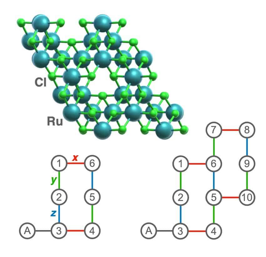
Figure showing a ball-and-stick model of ruthenium trichloride and two diagrams showing how the atoms' interactions are encoded into qubits. One diagram contains 10 numbered qubits, the other contains six

The quantum nature of complex molecules and materials makes them very challenging to simulate. To learn about their properties, a classical computer must store and process huge amounts of data.  Quantum computers bypass this by manipulating quantum systems directly, which in theory gives them an advantage over their classical counterparts. In practice, however, today’s quantum devices are sensitive to noise due to interactions with their environment, greatly diminishing their potential advantages.

In recent years, several teams (including Google in 2019 and researchers at the University of Science and Technology of China in 2020) have claimed that their quantum devices have “quantum advantage” over classical ones. However, the design of such experiments has largely played to the strengths of the quantum technologies in question rather than focusing on practical applications. This has made it difficult to assess how quantum devices would fare when applied to problems that are considered “useful” and intractable to classical computers, such as the simulation of complex quantum chemistry.

A team led by Garnet Chan of the California Institute of Technology, US, has now provided some insight into this question by performing simulations of two quantum chemistry problems on Google’s 53-qubit Weber quantum processor. The first simulation centred on a cluster of eight atoms inside the enzyme nitrogenase. This enzyme is an important component in a chemical process called nitrogen fixation, and a better understanding of its chemical properties could revolutionize fertilizer manufacturing. The second simulation focused on α-ruthenium trichloride, a material that may exist in an exotic quantum phase known as a “spin liquid” at low temperatures. Such materials are not well-understood and could have applications in data storage, topological quantum computation and even high-temperature superconductivity.

A long way to go

To calculate properties of interest in complex molecules and materials (such as their electronic energy states or low-energy excitations), physicists begin by mapping the electron spins of the atoms to qubits in the quantum device. The interactions between the electrons in the original chemical systems can then be captured by applying quantum “logic gates” in a certain order to the qubits. Finally, researchers extract information about the system by measuring the qubits and analysing the measurement outcomes. The more logic gates that need to be applied to capture the behaviour of the electron spins, the more noise and errors accumulate during the computation, leading to less reliable results.

Diagram of iron (pink) and sulphur (yellow) in a cluster within the nitrogenase enzyme. Two different clusters are shown, one large and one small, and each is represented by a diagram of qubit connectivity

In the new work, which is described in PRX Quantum, Chan and colleagues found that as their computations grew larger and required more gates, noise in the system quickly overwhelmed the useful information they wanted to extract.  For example, their experiments showed that while simulations of a crystalline lattice of α-ruthenium trichloride containing six atoms provided several meaningful results, this was no longer true when the size of the problem increased to 10 atoms or more.

Similarly, they could predict energy spectra for an 8-atom cluster found in nitrogenase reasonably well, but only after applying a myriad of post-processing techniques on the measurement data. What is more, the quantum experiments treated only simplified models of these systems, where classical computers are still able to provide exact estimates. This implies that quantum advantage is not yet a reality for such simulations.

Choosing the right problems

The results weren’t all bad news for quantum simulators, however. The researchers note that the capabilities of the quantum device depend dramatically on the type of problem being investigated, and for this series of experiments, they deliberately avoided choosing problems that catered to the Weber architecture. Instead, they chose to focus on how interesting the results of the simulation would be to the wider scientific community. When they modified their simulation parameters to better suit the quantum processor, they cut their computational resources in half, and therefore generated more meaningful results.

Ultimately, though, the results show that quantum devices that can replace classical supercomputers are still some ways off. “These overly simplified models of realistic chemical systems and materials that we chose to simulate in our work can be trivially simulated on a classical computer or even a personal laptop,” says Ruslan Tazhigulov, who led the study as a postdoc in Chan’s team and is now a data scientist at the pharmaceuticals firm EQRx. “After applying various error mitigation techniques, we demonstrate that there is still a long way to go for quantum devices to become practical tools in solving complex quantum chemistry problems.”

Competing pacemakers create distinctive triplets in heartbeats

A simple set of mathematical rules that accurately identifies signs of competing electrical signals in the heart has been developed by Leon Glass at McGill University and colleagues. The Canadian team did in vitro experiments that showed how abnormal beating triggered by anomalous electrical impulses can generate distinctive triplets. These triplets also appear in cardiac data from real patients and emerge from a simple mathematical model.

Our heartbeat is controlled by a cluster of specialized cells called a pacemaker. Located in the right atrium, the pacemaker broadcasts regular electrical “sinus” signals that cause heart muscle cells to contract in a regular manner. However, the heart can also contain an abnormal “ectopic” pacemaker located in a different part of the organ. This pacemaker can send out electrical signals, often at lower frequencies than the sinus pacemaker. The heart muscles can respond to both sinus and ectopic signals, leading to an irregular beating of the heart called ectopic arrhythmia.

To monitor for signs of ectopic pacemakers, scientists have developed mathematical models that predict the patterns of irregular heart activity that can emerge from this interference. In this new study, Glass’s team did experiments that put these models to the test.

Genetically modified

The team began by creating artificial pacemakers in cardiac cells taken from mice. These cells had been genetically modified so that the voltages produced by their cell membranes can be controlled using light. In thin layers of heart cells, the researchers used short light pulses to create pairs of pacemaker signals with slightly different frequencies (one sinus and the other ectopic), at spots positioned 8.5 mm apart.

In the region between the spots, they measured the number of sinus pulses detected between successive ectopic pulses – where the ectopic pulses occur at a lower frequency than the sinus pulses. Interference between the pulses causes this number to vary according to when and where in the sample the measurements are made.

Glass and colleagues discovered that the number of these intervening sinus pulses always came in distinctive triplets: such as 1, 2, 4; 1, 4, 6; or 4, 6, 11. These triplets all seemed to follow just two simple rules: that one number must be odd, and that the largest number must be one larger than the sum of the two smaller numbers.

Based on these rules, the researchers reproduced the same patterns in simple mathematical models, and also searched for the patterns in real electrocardiogram data from 47 patients. Despite the immense complexity of the heart, they detected the distinctive triplets in eight electrocardiograms.

Glass’s team hopes that the remarkable simplicity of the triplet pattern should make it easily detectable using wearable heartbeat monitors. This could strengthen the ability of cardiologists to diagnose and treat potentially life-threatening irregular heartbeats.

The research is described in Physical Review Letters.

Using radar to detect cosmic neutrinos in ice sheets, why Leo Szilard changed his mind on nuclear weapons

This episode of the Physics World Weekly podcast features Steven Prohira, who is co-leader of the Radar Echo Telescope collaboration, which aims to detect high energy cosmic neutrinos by sending radar waves through an Antarctic ice sheet. Based at the University of Kansas, Prohira explains the physics behind the project and talks about the fascinating history of previous attempts to use radar to detect particles from outer space.

Also on hand is Physics World’s Matin Durrani, who chats about the remarkable life of the Hungarian-American physicist Leo Szilard – who encouraged the US to develop nuclear weapons during the Second World War, but later opposed their use.

Robotic system uses multispectral imaging and artificial intelligence to search for earthquake victims

Robotic search system

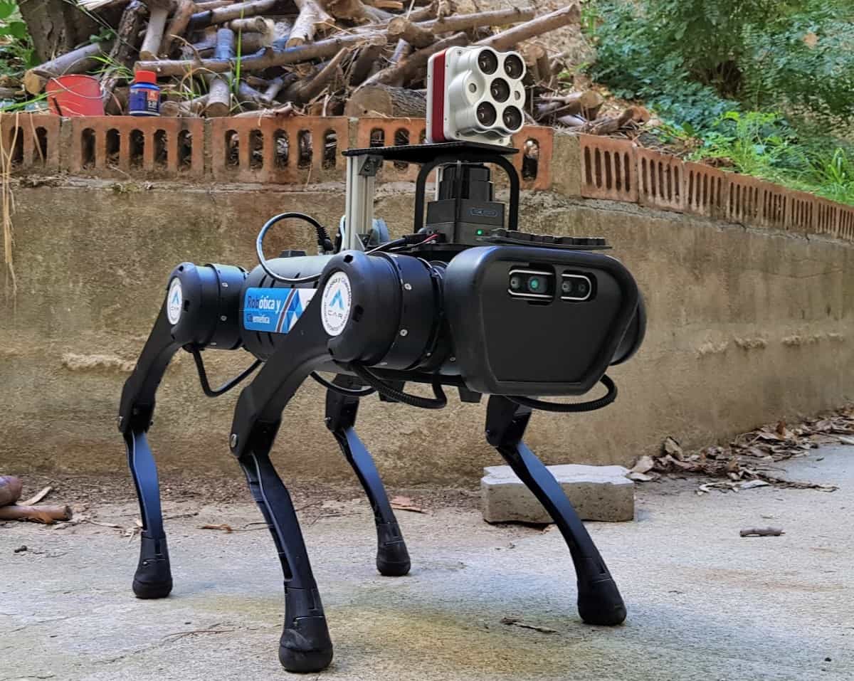
The horrific aftermath of the recent earthquakes that struck Syria and Turkey makes it clear why a range of technologies are needed to search for survivors in the rubble of destroyed buildings. Now, researchers at the Polytechnic University of Madrid have developed an autonomous robotic system that combines artificial intelligence with multispectral imaging to search for people in dangerous and difficult to access post-disaster environments.

The system uses an Altum multispectral camera that operates at five wavelengths – red, green and blue visible light; near infrared; and red edge light. The camera is attached to a remote-controlled quadruped robot that can be deployed in dangerous environments such as a collapsed building.

Christyan Cruz Ulloa and colleagues developed multispectral indexes that can be used to identify people within rubble. An index is a way of combining images taken at different wavelengths to enhance the ability of the multispectral imaging system to identify specific objects. Such indexes are often used to identify vegetation in satellite images, but no index had existed for identifying people in a rubble environment.

Convolutional neural networks

Once the team had identified an index that was suitable for both indoor and outdoor identification of people within a background of rubble, the next step was to train several convolutional neural networks (CNNs) to identify human forms – such as a torso, head and hand – within rubble. The researchers then compared efficacy of the different CNNs. This allowed them to select the best CNN for this application – a CNN called YOLOv5m.

The researchers then compared their prototype technique with previous systems, which tend to use either red-green-blue or infrared imaging to locate victims. They conclude that their multispectral system has greater versatility when it comes to locating people in both indoor and outdoor environments.

The team describes the system in a paper that has been accepted for publication in Machine Learning: Science and Technology.

Could next-generation perovskite detectors improve clinical X-ray imaging?

Perovskite-based X-ray detectors have a lot to offer the field of diagnostic imaging: low production costs, direct conversion, high absorption efficiency and superior spatial resolution to existing detectors. But while these advantages have been demonstrated in previous studies, researchers have not yet determined whether they translate to improvements in clinical applications.

To investigate this potential in more depth, researchers in the X-ray Cancer Imaging and Therapy Experimental (XCITE) Lab at the University of Victoria in Canada have performed virtual clinical trials on next-generation perovskite detectors integrated into common X-ray imaging devices, reporting their findings in Physics in Medicine & Biology.

The team investigated the perovskite crystal methylammonium lead bromide (MAPbBr3), which combines high charge carrier mobility and long carrier lifetimes, making it extremely sensitive to incident X-ray photons. Indeed, some MAPbBr3 crystals show equivalent performance to that of cadmium zinc telluride (CZT), a promising material used in cutting edge medical imaging techniques such as photon-counting CT.

To determine which imaging applications may suit perovskite detectors, the researchers used TOPAS Monte Carlo (MC) simulations to calculate the energy deposition efficiency (EDE, the fraction of absorbed energy relative to the incident energy) of MAPbBr3, for crystal thicknesses between 40 and 15 mm and beam energies from 20 keV to 6 MeV.

They compared the results with four other detector materials: amorphous selenium (a-Se), commonly used for mammography; caesium iodide (CsI), the standard detector material for kilovoltage (kV) CT; gadolinium oxysulphide (GOS), as used in kV and megavoltage (MV) imaging; and CZT.

Due to the lead content in MAPbBr3, the perovskite exhibited the highest energy absorption of all the detectors in the mammographic energy range. For MV imaging, only CZT had superior EDE, while for kV imaging, perovskite did not generally perform as well as the others. Based on these findings, the team chose three imaging systems to study: the Koning dedicated breast CT scanner, and Varian’s Truebeam kV and MV cone-beam CT (CBCT) systems.

“The EDE simulations motivated the inclusion of breast CT, a more niche imaging system that we would not have simulated otherwise,” explains first author Jericho O’Connell. “The kV- and MV-CBCT systems would have been included regardless, as they are key parts of the radiotherapy workflow.”

Virtual clinical trials

O’Connell and colleagues used Fastcat hybrid MC simulations to optimize the perovskite detector design for each application. By maximizing the detective quantum efficiency (DQE, the efficiency of converting an input signal to an output image), they calculated the optimal thicknesses for the perovskite crystals as 0.30, 0.86 and 1.99 mm, for breast CT, kV- and MV-CBCT, respectively. They then used these device-specific detectors in a series of virtual clinical trials.

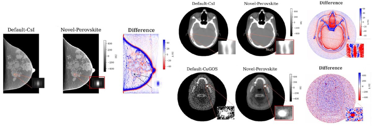
Images of phantoms using default detectors and perovskite detectors

For the breast CT trial, the researchers simulated a breast phantom with microcalcifications imaged using the default CsI detector and a perovskite detector with the same pixel pitch (0.194 mm). The perovskite detector increased contrast in the microcalcifications by 87%, clearly visualizing a calcified lesion that was poorly defined using the CsI detector. This could enable more accurate identification of such structures in breast cancer screening when using a perovskite detector, which can be manufactured at lower cost than CsI.

In the kV and MV CBCT virtual trials, the researchers imaged an XCAT head phantom. In both cases, the perovskite detector dramatically improved image quality compared with the default detectors. In the kV images, spatial resolution in fine bone features and tissue contrast was improved dramatically using the perovskite detector, increasing the CNR in brain and skull by 8% and 13%, respectively, compared with the CsI detector.

The MV image focused on a skull region containing silver fillings that would generally produce large streaking artefacts in kV images. The high efficiency of the perovskite detector compared with a GOS detector resulted in dramatic improvement in CNR and enabled a metal artefact-free image of the jaw. The researchers point out that the improved contrast in MV-CBCT images with a perovskite detector could enable imaging of patients on radiotherapy machines without a kV on-board imager, as is the case for most systems in low- and middle-income countries.

Replacing the current detectors on the breast CT, kV-CBCT and MV-CBCT machines with optimized perovskite detectors improved the DQE of these systems by 12.1%, 9.5% and 86.1%, respectively. “Perovskite detectors perform better than current detectors in breast CT and kV-CBCT applications, and are far superior to current MV-CBCT detectors in terms of CNR and DQE,” the researchers conclude.

Next, the team plans to create prototype perovskite-based flat-panel detectors to experimentally verify the virtual trials. “We are excited to report that we have sent off crystals to get a prototype pixelated detector made through AY Sensors,” O’Connell tells Physics World. “Stay tuned for the experimental characterization of the prototype detector.”

Science elevated to cabinet position in UK government shake-up

Science has been elevated to a cabinet-level position in the UK government for the first time in decades. The move occurred yesterday after Conservative prime minister Rishi Sunak announced the creation of a new Department for Science, Innovation and Technology, which will be led by Chippenham MP Michelle Donelan.

The new high profile for science came as part of a reshuffle of UK government, which will see the Department for Business, Energy and Industrial Strategy (BEIS) split into three separate departments. In addition to science, there is a new Department for Energy Security and Net Zero as well as a Department for Business and Trade.

Donelan, who was previously secretary of state for digital, culture, media and sport, has been made secretary of state for science, innovation and technology. George Freeman retains his science brief, becoming minister of state in the new department. He was previously minister of state for BEIS.

According to the UK government, the new science department will “focus on positioning the UK at the forefront of global scientific and technological advancement” and “direct record levels of research and development”. In a statement, it said that a department focused on turning “scientific and technical innovations into practical, appliable solutions will help make sure the UK is the most innovative economy in the world”.

‘Good news’

Tom Grinyer, group chief executive officer of the Institute of Physics, which publishes Physics World, says that the cabinet seat for science is good news for the UK as it “puts science and innovation exactly where they should be – right at the heart of government”.

Grinyer says that Donelan must work with the scientific community and other departments to ensure the government’s plan for the UK to become a global science superpower stays on track and “that there is a genuinely joined-up approach to science across government”.

Adrian Smith, president of the Royal Society, has also welcomed the creation of the new science department. In a statement, he says it signals that “research and innovation sit at the heart of the prime minister’s productivity and growth agenda”.

Smith believes that Donelan’s first job must be for the UK to secure association to Horizon Europe and other European Union (EU) science programmes. Participation in Horizon Europe had been agreed as part of the post-Brexit trade deal between the UK and EU, but the association agreement has not yet been signed and has since become a bargaining chip in other political issues related to Brexit.

“These schemes support outstanding international collaboration and without being part of them we are undermining the prime  minister’s stated ambition for the UK to be at the forefront of science and technology globally,” Smith says.

Daniel Rathbone, assistant director of the Campaign for Science and Engineering, also highlights access to European research programmes as a key issue for the new department, along with reform of the R&D tax-relief system.

Rathbone warns, however, that it will be vital that “the practicalities of making changes in Whitehall aren’t allowed to take away from the time and resources needed to drive forward the promising [science and innovation] agenda the government has previously set out”.

  • Businesses are being held back by a lack of apprentices in key physics-related roles. That is according to a new report by the Institute of Physics, which highlights a massive shortfall in the numbers of women pursuing such apprenticeships. The report, which surveyed nearly 300 apprentices and interviewed over 90 organisations, also finds that people are concerned about the cost of living crisis while employers say they struggle to get into schools to talk to young people about their options.

Designing materials and systems for decarbonizing chemicals and water industries

Want to learn more on this subject?

Storing cheap renewable electricity into chemical bonds (such as chemical energy storage) could be a transformative opportunity for long-duration energy storage that can address the intermittency of renewables and balance the mismatch between supply and demand at the grid.

The chemical industry primarily consumes fossil feedstock as an energy source, which has been the standard for more than a century. A paradigm shift is required to move towards a more sustainable route for chemical synthesis by electrifying and decarbonizing the modern chemical industry. As renewable electricity costs continue to decrease, there is a growing interest in fuels and chemical electrosynthesis.

This webinar focuses on developing systems, catalysts, and processes to use renewable electricity as an energetic driving force to produce high-value and high-energy molecules that can be utilized as either fuel, energy storers, and/or chemicals. Also discussed are future directions and strategies to manage the carbon, nitrogen, and water cycles, enabling a circular economy, mitigating waste, and promoting sustainability with positive social and environmental impacts through waste-water treatment and paving the way for nutrient recovery and recycling.

This research aims to develop next-generation sustainable industrial systems and processes that can aid in the transition to a net-zero-emission energy system and in meeting our world’s growing clean energy and water demands.

An interactive Q&A session follows the presentation.

Want to learn more on this subject?

Reza Nazemi is assistant professor in the Department of Mechanical Engineering and the School of Advanced Materials Discovery at Colorado State University (CSU). His group at CSU focuses on designing and developing systems and materials for clean-energy generation and waste-water treatment. In addition, his group leverages advanced spectroscopic and microscopic techniques to gain a mechanistic understanding of photoelectrochemical reactions for sustainable fuel and fertilizer production.

Dr Nazemi earned his PhD from the Woodruff School of Mechanical Engineering at the Georgia Institute of Technology (Georgia Tech) in 2020 followed by a postdoctoral fellowship there in the School of Chemistry and Biochemistry (2020–2021). From 2021–2022, he was a postdoctoral associate in the Department of Chemical and Environmental Engineering at Yale University. Reza’s notable recognition includes the 2022 ECS Colin Garfield Fink Fellowship, 2021 American Chemical Society Physical Chemistry Division Young Investigator Award, and 2018 Amazon Catalyst at ECS award. In 2020, he was the Georgia Tech Technology Innovation: Generating Economic Results Class of 2020 Fellow, and received the Georgia Research Alliance Award and Georgia Tech-Oak Ridge National Lab Seed Grant Award.


Practical approach to MR-guided online adaptive radiation therapy

Want to learn more on this subject?

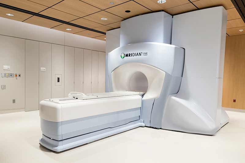
From Laura Bassi to Marie Curie, for centuries, women have been making important contributions to the world of physics. Now with ViewRay’s MRIdian system, women are leading the charge in bringing the latest advancement of MRI-guided radiation therapy to the forefront of radiation oncology and expanding the medical physics landscape.

MR-guided radiation therapy has stimulated a paradigm shift in external beam radiation therapy. Daily online adaptive therapy is now possible by introducing an advanced imaging modality during treatment delivery. Efforts to streamline its approach are prudent to broaden its availability and realize the clinical benefits. The implementation of a practical approach to an MRgRT online ART workflow will be discussed.

This series of five webinars will specifically highlight women physicists across the globe that are using MRIdian to transform cancer care as we know it. Jennifer Dolan of Henry Ford Health will present this webinar.

This presentation is the third in a series of Women in Medical Physics, supported by ViewRay.

Want to learn more on this subject?

Jennifer Dolan is the director of MR-guided radiation therapy physics at the Henry Ford Cancer Institute in Detroit, Michigan, where she also serves on the radiosurgery physics team. Dr Dolan received her PhD in nuclear engineering and radiological sciences from the University of Michigan in 2013. Upon completion of her studies, Jennifer worked in the nuclear security and nuclear well logging fields. She returned to the University of Michigan in 2016 to obtain her certificate in medical physics and completed her residency at Henry Ford Health in 2019. Her research interests focus on MR-guided adaptive radiation therapy and innovations in novel MR-guided applications to radiation therapy.

Dose tracking during radiotherapy could enable safer cancer treatments

Real-time dose monitoring

Radiotherapy of moving lesions is challenging. Delivery of the therapeutic radiation to a planned target volume may be impacted by organ motion, while anatomical deformations and set-up uncertainties can cause targeting errors. If radiation oncologists had an accurate, real-time 3D radiation dose distribution map, they would be able to alter the level or trajectory of radiation online to achieve more effective and safer treatments.

Ionizing radiation acoustic imaging (iRAI) is a non-invasive technology that could provide this capability. By reconstructing radiation dose using acoustic waves, iRAI can map dose deposition in tumours and adjacent healthy tissues and monitor dose accumulation in real time during radiotherapy, without the need to use additional radiation sources.

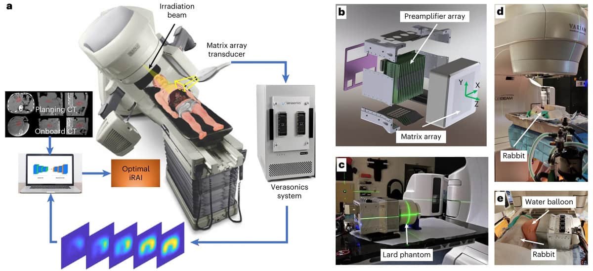
A multi-speciality research team at the University of Michigan and Moffitt Cancer Center has now developed a clinical grade iRAI volumetric imaging system. The system, described in Nature Biotechnology, achieved 3D semiquantitative mapping of X-ray beam delivery deep into the body during radiotherapy of a patient with liver metastases.

The iRAI technique works via the thermoacoustic effect. When a high-energy pulsed photon beam generated by a linear accelerator strikes body tissue, it is absorbed. This absorbed energy transfers into heat, which causes localized thermal expansion and generates acoustic waves. These waves are weak, however, and usually undetectable by clinical ultrasound technology.

The new iRAI system detects the acoustic signals with a custom-designed 2D matrix array transducer and a matching multi-channel preamplifier board, driven by a commercial research ultrasound system. The amplified signal is then transferred into an ultrasound device to construct dose-related images in real time.

The researchers explain that their dual-modality system, which combines iRAI with ultrasound imaging, offers “a promising solution to solve the need for real-time monitoring of beam position and online assessment of delivering dose during radiotherapy”. The ultrasound image presents the morphological tissue structures and motion in the body, as well as functional information such as blood flow and vascular density, while the iRAI image can map and quantify the spatially distributed dose deposition in different biological tissues.

“This clinical trial was a pilot study to assess the feasibility of using iRAI in patients receiving abdominal stereotactic body radiation therapy (SBRT),” explains clinical principal investigator Kyle Cuneo from Michigan’s Rogel Cancer Center. “Its findings are enabling us to optimize the iRAI system.”

iRAI experimental setups

For their proof-of-concept study, the researchers validated the system in a cylindrical lard phantom, a rabbit and then a patient undergoing abdominal SBRT. To enhance the signal-to-noise ratio (SNR) when detecting radiation acoustic signals, they selected a central frequency of 0.35 MHz to match the power spectrum of the acoustic signals generated by the 4 µs X-ray pulse. The SNR was further enhanced by the 1024-channel preamplifier with 46 dB gain integrated with the 2D matrix array, and by displaying the iRAI images with 25 times averaging.

After verifying the system performance using the phantom, the team created and tested a clinical treatment plan to irradiate the liver of a rabbit. The iRAI measurements showed high consistency between the measured dose distribution and that generated by the treatment planning system.

The team then prepared radiotherapy plans for the study participant, with the treatment plan for each fraction divided into two parts. The first part was for iRAI imaging and comprised 2.087 and 0.877 Gy beams delivered in the superior and inferior anterior directions, respectively. This was followed by a volumetric-modulated arc therapy plan (without iRAI imaging) to ensure that the total delivered radiation dose met the clinical requirements.

Both the dose locations and the overall distributions of iRAI measurements matched well with the treatment plan. The iRAI volumetric imaging was able to map the high-dose area with high accuracy. The researchers note that they need to optimize the mapping accuracy for lower dose intensity areas, improve spatial resolution, and develop a comprehensive calibration protocol to provide absolute dose measurement, using advanced reconstruction techniques exploiting artificial intelligence.

Grant principal investigator Issam El Naqa from the Moffitt Cancer Center advises that the current system will be augmented with real-time ultrasound imaging and will also be evaluated in the context of high-risk delivery scenarios such as FLASH radiotherapy.

“One potential application of this technology in the future is real-time adaptive treatment delivery. Current adaptive treatment techniques are based mainly on anatomical changes in the tumour and organs-at-risk (OARs),” explains Cuneo. “With iRAI, we can use both anatomical information, and more importantly, dosimetric information to adapt the radiation plan. This could allow for dose escalation in the target, especially in situations where there is an adjacent OAR, and provide for safer treatments by accurately quantifying the true dose delivered to the target and OARs during each fraction.”

“The system has the unique capability of visualizing radiation deposition while monitoring organ motion, allowing better pinpointing of radiation to targeted tumours while sparing uninvolved tissue in a cost saving manner,” El Naqa adds. “This can be equally applied in both developed and developing countries where financial resources are scarce, leading to improved patient care and better outcomes in these places.”

Copyright © 2026 by IOP Publishing Ltd and individual contributors